File:Sabin1901 plate02.jpg
Original file (1,723 × 1,000 pixels, file size: 590 KB, MIME type: image/jpeg)
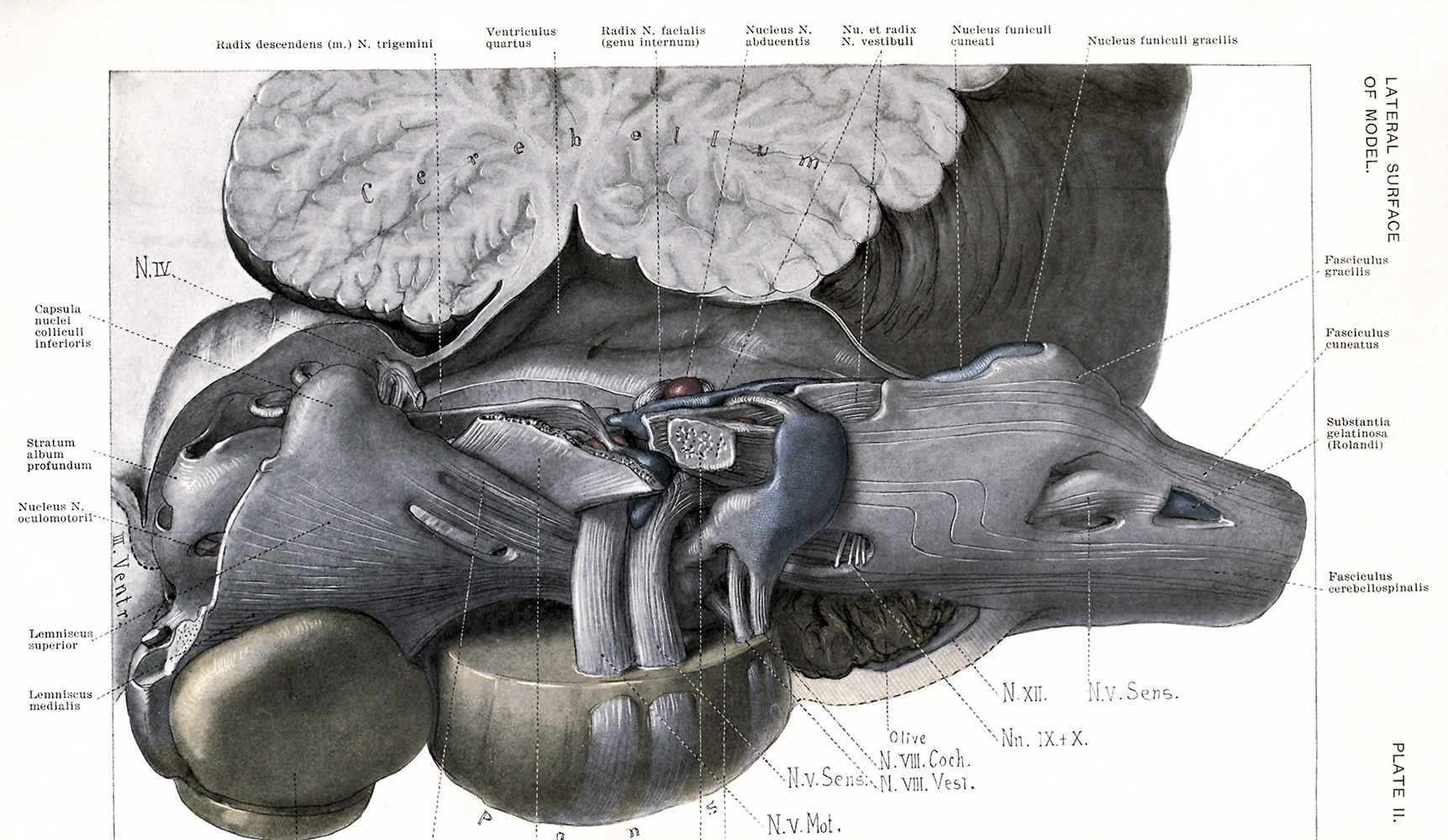
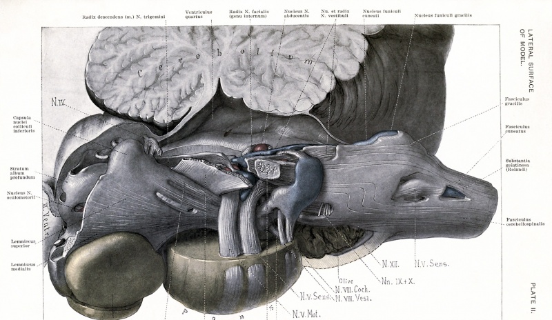
Plate II. View of the model from the lateral surface
This view is designed to relate the model to the cord, the cerebellum and the cerebrum. The cut edge of the cord shows on the extreme right. The following points will make the position of the model clear: the dorsal and lateral funiculi and the dorsal horn of the spinal cord, the cerebellum, the fourth ventricle, the inferior and superior colliculi and the third ventricle.
The color system is as follows: all fibres are in white and black, all nuclei in colors. Red represents the nuclei of the motor cerebral nerves, blue the nuclei of the sensory cerebral nerves and yellow all other nuclei.
Nu. et Radix N. vestibuli: The nucleus is distinguishable from the root by its color. The ascending and descending parts of the root are to be determined by their relation to the entering root-bundle of the nerve. The part of the vestibular nucleus distal to the nucleus N. abducentis is the nucleus N. vestibuli medialis; the part proximal, is the nucleus N. vestibuli superior. The nucleus N. vestibuli lateralis (Deiters'), (pars lateralis) lies in the vestibular tract just dorsal to the corpus restiforme.
| Historic Disclaimer - information about historic embryology pages |
|---|
 |
- Figures: Plate 1 | Plate 2 | Plate 3 | Plate 4 | Plate 5 | Plate 6 | Plate 7 | Plate 8 | Fig. 52. Key to Planes of Sections
Reference
Sabin FR. and Knower H. An atlas of the medulla and midbrain, a laboratory manual (1901) Baltimore: Friedenwald.
Cite this page: Hill, M.A. (2024, April 23) Embryology Sabin1901 plate02.jpg. Retrieved from https://embryology.med.unsw.edu.au/embryology/index.php/File:Sabin1901_plate02.jpg
- © Dr Mark Hill 2024, UNSW Embryology ISBN: 978 0 7334 2609 4 - UNSW CRICOS Provider Code No. 00098G
File history
Click on a date/time to view the file as it appeared at that time.
| Date/Time | Thumbnail | Dimensions | User | Comment | |
|---|---|---|---|---|---|
| current | 15:50, 9 June 2015 | | 1,723 × 1,000 (590 KB) | Z8600021 (talk | contribs) |
You cannot overwrite this file.
File usage
The following page uses this file: