File:Rugh 139.jpg: Difference between revisions
mNo edit summary |
mNo edit summary |
||
| (One intermediate revision by the same user not shown) | |||
| Line 1: | Line 1: | ||
==Early development of the thyroid gland of the frog== | |||
(A,B) Separation of the thyroid anlage from the pharynx. | |||
(C) Division of the thyroid anlage into lobes at the 9 mm stage. | |||
(D) Sagittal section of the 11 mm stage showing the position of the thyroid anlage in relation to the pharynx. | |||
---- | |||
The thyroid is an endocrine gland which arises as a single median thickening and evagination from the floor of the pharynx between the base of the second pair of visceral arches, just before the time of hatching and at about the 5 mm. stage. It later becomes a bi-lobed and solid organ, far removed from its site of origin. Only its original lining is endodermal, the bulk of the gland being mesodermal in origin. At about the 10 mm. stage, however, it becomes separated from the pharyngeal floor as a closed sac. This divides into two lobular and vesicular masses, and moves to a position on either side of the hyoid cartilage apparatus. The thyroid gland of the 15 mm. stage is divided but retains a connection at its anterior ends by a short isthmus, so that it takes the shape of an inverted "Y," with a short base. The wings move posteriorly, along the ventral face of the hyoid cartilage, and these changes are correlated with gradual changes in the hyoid apparatus. The glands enlarge, the surrounding cartilaginous elements expand, the major blood vessels become enmeshed in them, and eventually they come to lie close to the heart. At the forelimb emergence stage of metamorphosis there is a marked distension of the follicles, and by the tail resorption stage there is high epithelium liquefaction while erosion of the colloid and follicular collapse occur. At this time the genio-hyoid, hypoglossus, andsternohyoid muscles are in close proximity to the thyroid, a situation which does not persist to the adult. The gland shows hyperactivity during metamorphosis with relative inactivity at the completion of the metamorphic process. Its activity during these phases of development is closely correlated with the development and function of the pituitary gland, particularly of the basophilic cells in the pituitary gland. In later stages it may be seen attached to the ventral aspect of the hypoglossus muscle. The fully formed thyroid gland consists of separate follicles, each made up of a single circular layer of cuboidal (endodermal) epithelial cells, in the center of which is a lumen filled with a colloidal mass. | |||
{{Rugh1951 footer}} | {{Rugh1951 footer}} | ||
Latest revision as of 08:11, 17 April 2013
Early development of the thyroid gland of the frog
(A,B) Separation of the thyroid anlage from the pharynx.
(C) Division of the thyroid anlage into lobes at the 9 mm stage.
(D) Sagittal section of the 11 mm stage showing the position of the thyroid anlage in relation to the pharynx.
The thyroid is an endocrine gland which arises as a single median thickening and evagination from the floor of the pharynx between the base of the second pair of visceral arches, just before the time of hatching and at about the 5 mm. stage. It later becomes a bi-lobed and solid organ, far removed from its site of origin. Only its original lining is endodermal, the bulk of the gland being mesodermal in origin. At about the 10 mm. stage, however, it becomes separated from the pharyngeal floor as a closed sac. This divides into two lobular and vesicular masses, and moves to a position on either side of the hyoid cartilage apparatus. The thyroid gland of the 15 mm. stage is divided but retains a connection at its anterior ends by a short isthmus, so that it takes the shape of an inverted "Y," with a short base. The wings move posteriorly, along the ventral face of the hyoid cartilage, and these changes are correlated with gradual changes in the hyoid apparatus. The glands enlarge, the surrounding cartilaginous elements expand, the major blood vessels become enmeshed in them, and eventually they come to lie close to the heart. At the forelimb emergence stage of metamorphosis there is a marked distension of the follicles, and by the tail resorption stage there is high epithelium liquefaction while erosion of the colloid and follicular collapse occur. At this time the genio-hyoid, hypoglossus, andsternohyoid muscles are in close proximity to the thyroid, a situation which does not persist to the adult. The gland shows hyperactivity during metamorphosis with relative inactivity at the completion of the metamorphic process. Its activity during these phases of development is closely correlated with the development and function of the pituitary gland, particularly of the basophilic cells in the pituitary gland. In later stages it may be seen attached to the ventral aspect of the hypoglossus muscle. The fully formed thyroid gland consists of separate follicles, each made up of a single circular layer of cuboidal (endodermal) epithelial cells, in the center of which is a lumen filled with a colloidal mass.
| Historic Disclaimer - information about historic embryology pages |
|---|
 |
Reference
Rugh R. Book - The Frog Its Reproduction and Development. (1951) The Blakiston Company.
Cite this page: Hill, M.A. (2024, April 16) Embryology Rugh 139.jpg. Retrieved from https://embryology.med.unsw.edu.au/embryology/index.php/File:Rugh_139.jpg
- © Dr Mark Hill 2024, UNSW Embryology ISBN: 978 0 7334 2609 4 - UNSW CRICOS Provider Code No. 00098G
File history
Click on a date/time to view the file as it appeared at that time.
| Date/Time | Thumbnail | Dimensions | User | Comment | |
|---|---|---|---|---|---|
| current | 19:40, 14 April 2013 | 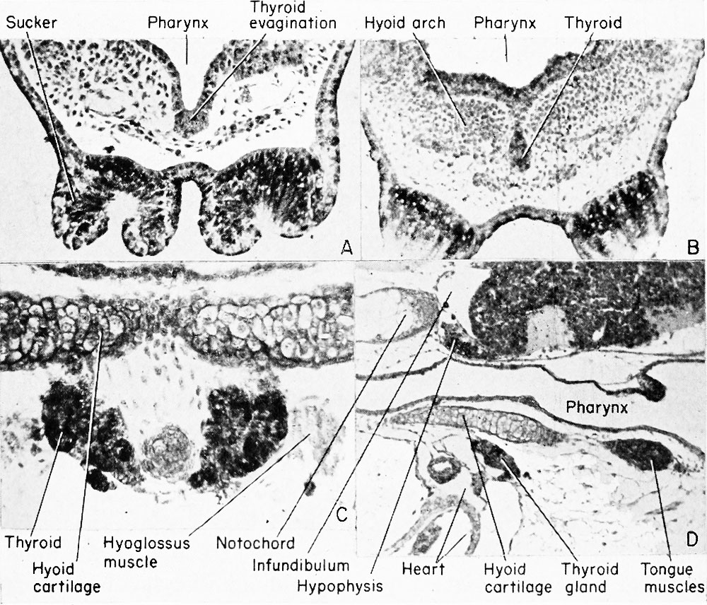 | 1,000 × 856 (261 KB) | Z8600021 (talk | contribs) |
You cannot overwrite this file.
File usage
The following 2 pages use this file: