File:Rugh 071.jpg: Difference between revisions
mNo edit summary |
mNo edit summary |
||
| Line 1: | Line 1: | ||
== | ==The process of gastrulation== | ||
(A) The blastula stage, prior to any gastrulation movement. | |||
(B) Movement of the blastula cells preliminary to gastrulation. | |||
(C) Blastoporal view of successive phases of gastrulation; (solid line) lip of blastopore, {dotted line) germ ring, to be subsequently incorporated into the blastoporal lips. | |||
(D) Lateral view of sagittal section during late gastrulation showing the origin of the mesial notochord, and the lateral mesoderm from the proliferated chorda-mesoderm cells at the dorsal lip. | |||
(E) Composite drawing to illustrate the germ layer relations in the later gastrula of the frog. The medullary plate (ectoderm) is not indicated; {alternate dots and dashes) notochord, {heavy stippling) notochord, {sparse stippling) mesoderm, (cellular markings) ectoderm. | |||
{{Rugh1951 footer}} | {{Rugh1951 footer}} | ||
[[Category:Gastrulation]] | |||
Revision as of 12:41, 12 April 2013
The process of gastrulation
(A) The blastula stage, prior to any gastrulation movement.
(B) Movement of the blastula cells preliminary to gastrulation.
(C) Blastoporal view of successive phases of gastrulation; (solid line) lip of blastopore, {dotted line) germ ring, to be subsequently incorporated into the blastoporal lips.
(D) Lateral view of sagittal section during late gastrulation showing the origin of the mesial notochord, and the lateral mesoderm from the proliferated chorda-mesoderm cells at the dorsal lip.
(E) Composite drawing to illustrate the germ layer relations in the later gastrula of the frog. The medullary plate (ectoderm) is not indicated; {alternate dots and dashes) notochord, {heavy stippling) notochord, {sparse stippling) mesoderm, (cellular markings) ectoderm.
| Historic Disclaimer - information about historic embryology pages |
|---|
 |
Reference
Rugh R. Book - The Frog Its Reproduction and Development. (1951) The Blakiston Company.
Cite this page: Hill, M.A. (2024, April 19) Embryology Rugh 071.jpg. Retrieved from https://embryology.med.unsw.edu.au/embryology/index.php/File:Rugh_071.jpg
- © Dr Mark Hill 2024, UNSW Embryology ISBN: 978 0 7334 2609 4 - UNSW CRICOS Provider Code No. 00098G
File history
Click on a date/time to view the file as it appeared at that time.
| Date/Time | Thumbnail | Dimensions | User | Comment | |
|---|---|---|---|---|---|
| current | 12:05, 12 April 2013 | 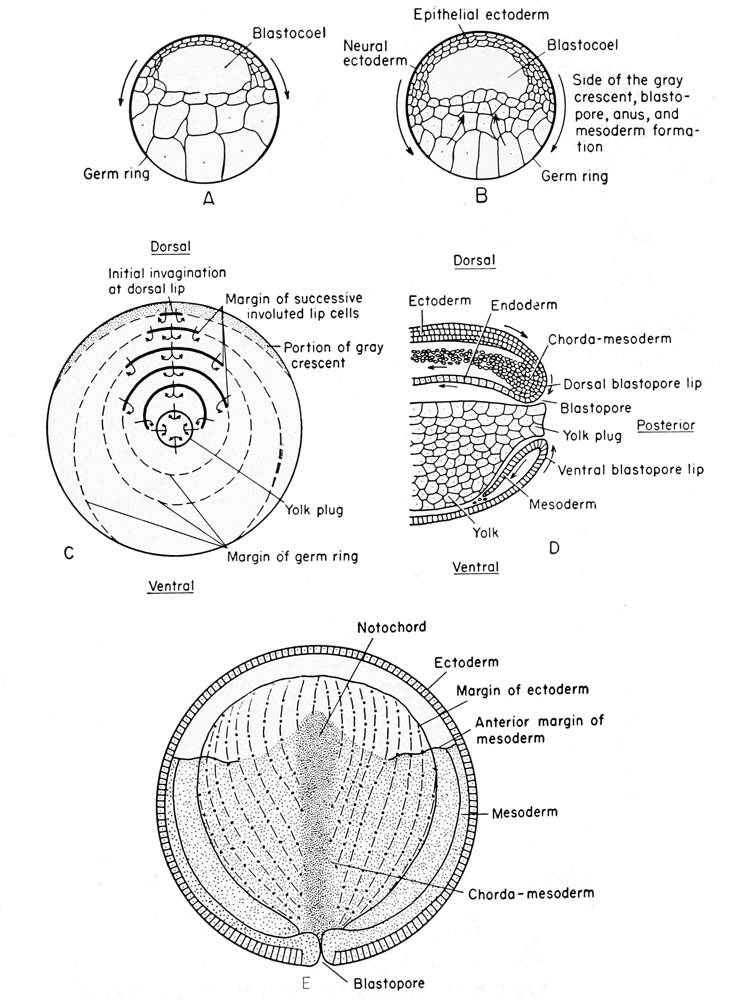 | 737 × 1,000 (140 KB) | Z8600021 (talk | contribs) |
You cannot overwrite this file.
File usage
The following 3 pages use this file: