File:Rugh 017.jpg
Original file (756 × 1,200 pixels, file size: 331 KB, MIME type: image/jpeg)
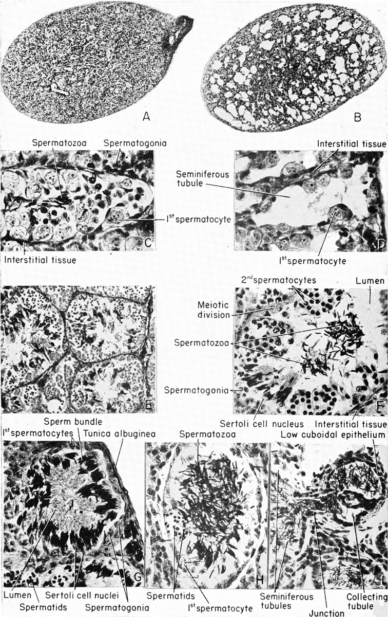
Spermatogenesis in the frog: Rana pipiens
(A) Testis of a recently metamorphosed frog.
(B) Similar to "A" except that the frog was previously treated with the anterior pituitary hormone to evacuate the seminiferous tubules, leaving only the interstitial tissue, spermatogonia, and a few primary spermatocytes. The post-breeding condition.
(C) High-power magnification of "A." (D) High-power magnification of "B."
(E) Testis of an August frog, showing all stages of spermatogenesis.
(F) Partially (pituitary) activated testis, similar to "E," showing released spermatozoa within the lumen, and all stages of spermatogenesis around the periphery of the seminiferous tubule.
(G) Testis of a hibernating, pre-breeding frog. Note the clusters of mature spermatozoa attached to single Sertoli cells. The lumen is filled with tails, few spermatogonia, and primary spermatocytes around the periphery of the seminiferous tubule.
(H) Testis of the male during amplexus, showing spermatozoa liberated into the lumen of the seminiferous tubule. (I) Collecting tubule of the frog testis full of mature spermatozoa, showing their origin from the connecting seminiferous tubules. Collecting tubules are lined with low cuboidal epithelium and are continuous with the vasa efferentia and the Malpighian corpuscles of the kidney.
| Historic Disclaimer - information about historic embryology pages |
|---|
 |
Reference
Rugh R. Book - The Frog Its Reproduction and Development. (1951) The Blakiston Company.
Cite this page: Hill, M.A. (2024, April 20) Embryology Rugh 017.jpg. Retrieved from https://embryology.med.unsw.edu.au/embryology/index.php/File:Rugh_017.jpg
- © Dr Mark Hill 2024, UNSW Embryology ISBN: 978 0 7334 2609 4 - UNSW CRICOS Provider Code No. 00098G
File history
Click on a date/time to view the file as it appeared at that time.
| Date/Time | Thumbnail | Dimensions | User | Comment | |
|---|---|---|---|---|---|
| current | 16:06, 6 April 2013 | | 756 × 1,200 (331 KB) | Z8600021 (talk | contribs) | {{Rugh1951 footer}} |
You cannot overwrite this file.
File usage
The following 2 pages use this file: