File:Retinal Disc to form Optic Cup at Carnegie stage 14.jpg
Original file (572 × 800 pixels, file size: 400 KB, MIME type: image/jpeg)
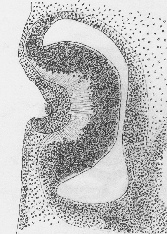
At Carnegie stage 14, the retinal disc becomes invaginated to form the optic cup. Lens Pit can also be seen forming. Contour forming slowly.
Based upon images by: R O'Rahilly The prenatal development of the human eye. Exp. Eye Res.: 1975, 21(2);93-112 Figure 3.
Beginning six months after publication, I, (z3374173) grant the public the non-exclusive right to copy, distribute, or display the Work under a Creative Commons Attribution-Noncommercial-Share Alike 3.0 Unported license, as described at http://creativecommons.org/licenses/by-nc-sa/3.0/ and http://creativecommons.org/licenses/by-nc-sa/3.0/legalcode.
File history
Click on a date/time to view the file as it appeared at that time.
| Date/Time | Thumbnail | Dimensions | User | Comment | |
|---|---|---|---|---|---|
| current | 21:11, 4 October 2012 | | 572 × 800 (400 KB) | Z3374173 (talk | contribs) | At Carnegie stage 14, the retinal disc becomes invaginated to form the optic cup. Lens Pit can also be seen forming. Contour forming slowly. Based upon images by: R O'Rahilly The prenatal development of the human eye. Exp. Eye Res.: 1975, 21(2);93-112 F |
You cannot overwrite this file.
File usage
The following 2 pages use this file: