File:Regions of the brain.jpg: Difference between revisions
No edit summary |
No edit summary |
||
| Line 1: | Line 1: | ||
Coronal view of the brain, showing the main basal ganglia nuclei. The section is angled rostrocaudally to encounter most of the BG nuclei in a single section. C, cortex; STR, striatum; GPe, globus pallidus pars externa; GPi, globus pallidus pars interna; Th, thalamus; STN, subthalamic nucleus; SNc, substantia nigra pars compacta; SNr, substantia nigra pars reticulata. | Coronal view of the brain, showing the main basal ganglia nuclei. The section is angled rostrocaudally to encounter most of the BG nuclei in a single section. '''C''', cortex; '''STR,''' striatum; '''GPe''', globus pallidus pars externa; '''GPi''', globus pallidus pars interna; '''Th''', thalamus; '''STN''', subthalamic nucleus; '''SNc''', substantia nigra pars compacta; '''SNr''', substantia nigra pars reticulata. | ||
<ref><pubmed>11909992</pubmed></ref> | <ref><pubmed>11909992</pubmed></ref> | ||
''Structures significant in HD, Striatum, Cortex & Thalamus.'' | |||
===Reference=== | ===Reference=== | ||
Revision as of 05:37, 11 October 2011
Coronal view of the brain, showing the main basal ganglia nuclei. The section is angled rostrocaudally to encounter most of the BG nuclei in a single section. C, cortex; STR, striatum; GPe, globus pallidus pars externa; GPi, globus pallidus pars interna; Th, thalamus; STN, subthalamic nucleus; SNc, substantia nigra pars compacta; SNr, substantia nigra pars reticulata. [1]
Structures significant in HD, Striatum, Cortex & Thalamus.
Reference
- ↑ <pubmed>11909992</pubmed>
http://physiologyonline.physiology.org/content/17/2/51.full
Organizations Requesting Permission to Reuse the Work of Others for Educational Purposes
APS grants permission for free use of our articles (in whole or in part) in educational materials provided
there is no charge or fee for those materials, and/or those materials are not directly or indirectly commercially supported. If a fee is charged or the materials are commercially supported, a fee will be assessed when permission is granted.
- Note - This image was originally uploaded as part of a student project and may contain inaccuracies in either description or acknowledgements. Students have been advised in writing concerning the reuse of content and may accidentally have misunderstood the original terms of use. If image reuse on this non-commercial educational site infringes your existing copyright, please contact the site editor for immediate removal.
Cite this page: Hill, M.A. (2024, April 24) Embryology Regions of the brain.jpg. Retrieved from https://embryology.med.unsw.edu.au/embryology/index.php/File:Regions_of_the_brain.jpg
- © Dr Mark Hill 2024, UNSW Embryology ISBN: 978 0 7334 2609 4 - UNSW CRICOS Provider Code No. 00098G
File history
Click on a date/time to view the file as it appeared at that time.
| Date/Time | Thumbnail | Dimensions | User | Comment | |
|---|---|---|---|---|---|
| current | 06:35, 10 October 2011 | 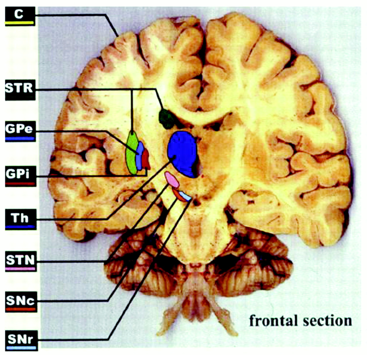 | 1,280 × 1,239 (348 KB) | Z3290270 (talk | contribs) | Coronal view of the brain, showing the main basal ganglia nuclei. The section is angled rostrocaudally to encounter most of the BG nuclei in a single section. C, cortex; STR, striatum; GPe, globus pallidus pars externa; GPi, globus pallidus pars interna; |
You cannot overwrite this file.
File usage
The following file is a duplicate of this file (more details):
There are no pages that use this file.