File:Regions of NC Cells.jpg
From Embryology

Size of this preview: 579 × 599 pixels. Other resolution: 1,460 × 1,511 pixels.
Original file (1,460 × 1,511 pixels, file size: 204 KB, MIME type: image/jpeg)
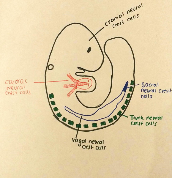
This image shows the regions where the different neural crest cells can be found. Note how these regions were discovered initially as the trunk and cranial neural crest cells, but later the existence of sacral, vagal and cardiac neural crest cells was confirmed. It is the cells that migrate vertically from the trunk neural crest that become the adrenal medulla.
This image was inspired by the figure 2 on the following page: https://jacobspublishers.com/molecular-mechanism-of-cranial-neural-crest-cell-development/
File history
Click on a date/time to view the file as it appeared at that time.
| Date/Time | Thumbnail | Dimensions | User | Comment | |
|---|---|---|---|---|---|
| current | 17:25, 7 October 2018 |  | 1,460 × 1,511 (204 KB) | Z5091101 (talk | contribs) | This image shows the regions where the different neural crest cells can be found. Note how these regions were discovered initially as the trunk and cranial neural crest cells, but later the existence of sacral, vagal and cardiac neural crest cells was... |
You cannot overwrite this file.
File usage
The following 2 pages use this file: