File:Reduced cardiomyocyte differentiation at the venous pole in isl1 mutant embryos.png: Difference between revisions
(Single z-scan of the atrium of a representative wild-type sibling (A-C) and mutant (D-F) embryo. White arrow (B) indicates eGFPposDsRedneg cardiomyocytes at the venous pole of the WT sibling heart; red arrow (E) indicates the venous pole of the isl1 mu...) |
No edit summary |
||
| Line 1: | Line 1: | ||
===Description=== | |||
''Figure 16 | |||
'' | |||
Single z-scan of the atrium of a representative wild-type sibling (A-C) and mutant (D-F) embryo. White arrow (B) indicates eGFPposDsRedneg cardiomyocytes at the venous pole of the WT sibling heart; red arrow (E) indicates the venous pole of the isl1 mutant where only very few eGFPposDsRedneg cells are present. (G,H) Single z-scan of a confocal image at the level of the arterial pole showing the eGFPposDsRedneg cardiomyocytes in the isl1 mutant (white arrow). V, ventricle: A, atrium. Scale bar: 50 μm. (I) Schematic of the location of the eGFPposDsRedpos cells (yellow) and the eGFPposDsRedneg (green) cells in the arterial pole of the isl1 mutant. (J,K) The number of eGFPposDsRedpos (J) and eGFPposDsRedneg (K) cardiomyocytes per embryo. (K) The eGFPposDsRedneg cardiomyocytes are subdivided into eGFPposDsRedneg cells being present at either the arterial or the venous pole. Note the significant reduction of eGFPposDsRedneg cells at the venous pole in the isl1 mutants. | Single z-scan of the atrium of a representative wild-type sibling (A-C) and mutant (D-F) embryo. White arrow (B) indicates eGFPposDsRedneg cardiomyocytes at the venous pole of the WT sibling heart; red arrow (E) indicates the venous pole of the isl1 mutant where only very few eGFPposDsRedneg cells are present. (G,H) Single z-scan of a confocal image at the level of the arterial pole showing the eGFPposDsRedneg cardiomyocytes in the isl1 mutant (white arrow). V, ventricle: A, atrium. Scale bar: 50 μm. (I) Schematic of the location of the eGFPposDsRedpos cells (yellow) and the eGFPposDsRedneg (green) cells in the arterial pole of the isl1 mutant. (J,K) The number of eGFPposDsRedpos (J) and eGFPposDsRedneg (K) cardiomyocytes per embryo. (K) The eGFPposDsRedneg cardiomyocytes are subdivided into eGFPposDsRedneg cells being present at either the arterial or the venous pole. Note the significant reduction of eGFPposDsRedneg cells at the venous pole in the isl1 mutants. | ||
Note that eGFPposDsRedneg represent cells that have initiated differentiation at a later stage in contrast to eGFPposDsRedpos which represent cells that have initiated differentiation early. | Note that eGFPposDsRedneg represent cells that have initiated differentiation at a later stage in contrast to eGFPposDsRedpos which represent cells that have initiated differentiation early. | ||
===Reference=== | ===Reference=== | ||
The image is taken from | The image is taken from <pubmed>19395641</pubmed> | ||
{{Template: Student Image}} | |||
===Copyright=== | |||
Copyright statement: Articles published in these journals are in the public domain and may be used and reproduced without special permission. However, anyone using the material is requested to properly cite and acknowledge the source. | |||
{{Template:Student Image}} | |||
Revision as of 21:50, 25 October 2017
Description
Figure 16
Single z-scan of the atrium of a representative wild-type sibling (A-C) and mutant (D-F) embryo. White arrow (B) indicates eGFPposDsRedneg cardiomyocytes at the venous pole of the WT sibling heart; red arrow (E) indicates the venous pole of the isl1 mutant where only very few eGFPposDsRedneg cells are present. (G,H) Single z-scan of a confocal image at the level of the arterial pole showing the eGFPposDsRedneg cardiomyocytes in the isl1 mutant (white arrow). V, ventricle: A, atrium. Scale bar: 50 μm. (I) Schematic of the location of the eGFPposDsRedpos cells (yellow) and the eGFPposDsRedneg (green) cells in the arterial pole of the isl1 mutant. (J,K) The number of eGFPposDsRedpos (J) and eGFPposDsRedneg (K) cardiomyocytes per embryo. (K) The eGFPposDsRedneg cardiomyocytes are subdivided into eGFPposDsRedneg cells being present at either the arterial or the venous pole. Note the significant reduction of eGFPposDsRedneg cells at the venous pole in the isl1 mutants. Note that eGFPposDsRedneg represent cells that have initiated differentiation at a later stage in contrast to eGFPposDsRedpos which represent cells that have initiated differentiation early.
Reference
The image is taken from <pubmed>19395641</pubmed>
Copyright
Copyright statement: Articles published in these journals are in the public domain and may be used and reproduced without special permission. However, anyone using the material is requested to properly cite and acknowledge the source.
- Note - This image was originally uploaded as part of an undergraduate science student project and may contain inaccuracies in either description or acknowledgements. Students have been advised in writing concerning the reuse of content and may accidentally have misunderstood the original terms of use. If image reuse on this non-commercial educational site infringes your existing copyright, please contact the site editor for immediate removal.
File history
Click on a date/time to view the file as it appeared at that time.
| Date/Time | Thumbnail | Dimensions | User | Comment | |
|---|---|---|---|---|---|
| current | 12:01, 20 November 2017 |  | 505 × 243 (76 KB) | Z8600021 (talk | contribs) | |
| 09:19, 24 October 2017 | 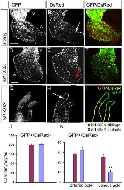 | 416 × 639 (280 KB) | Z5076466 (talk | contribs) | Single z-scan of the atrium of a representative wild-type sibling (A-C) and mutant (D-F) embryo. White arrow (B) indicates eGFPposDsRedneg cardiomyocytes at the venous pole of the WT sibling heart; red arrow (E) indicates the venous pole of the isl1 mu... |
You cannot overwrite this file.
File usage
The following file is a duplicate of this file (more details):
The following 2 pages use this file: