File:Ramsey1972 fig07.jpg
From Embryology
Size of this preview: 800 × 479 pixels. Other resolution: 1,280 × 767 pixels.
Original file (1,280 × 767 pixels, file size: 143 KB, MIME type: image/jpeg)
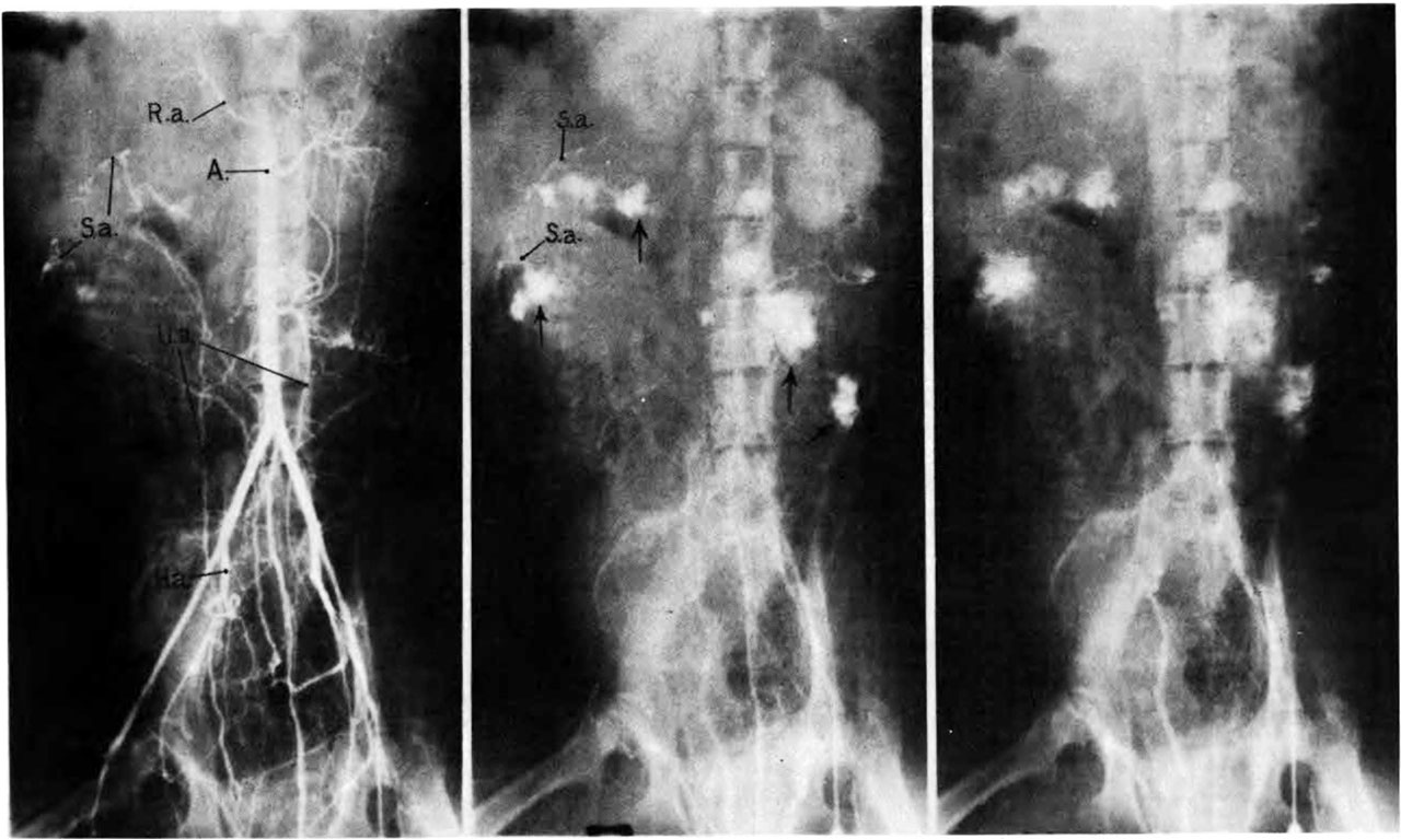
Fig. 7. Photographs of X rays femoral artery, endometrial spiral artery and intervillous space
Made at 2, 3, and 7% seconds respectively after injection of contrast material into a femoral artery of a monkey. (R.a.) renal artery; (S.a.) endometrial spiral artery;—>“spurts” into intervillous space. Carnegie Collection Monkey 60/14, 100th day of pregnancy.
(Reprinted with permission from Ramsey, er al. Montanino Editore, Napoli. ll: 1779-1784, 1962.)
File history
Click on a date/time to view the file as it appeared at that time.
| Date/Time | Thumbnail | Dimensions | User | Comment | |
|---|---|---|---|---|---|
| current | 07:06, 23 February 2017 | | 1,280 × 767 (143 KB) | Z8600021 (talk | contribs) | |
| 07:06, 23 February 2017 | 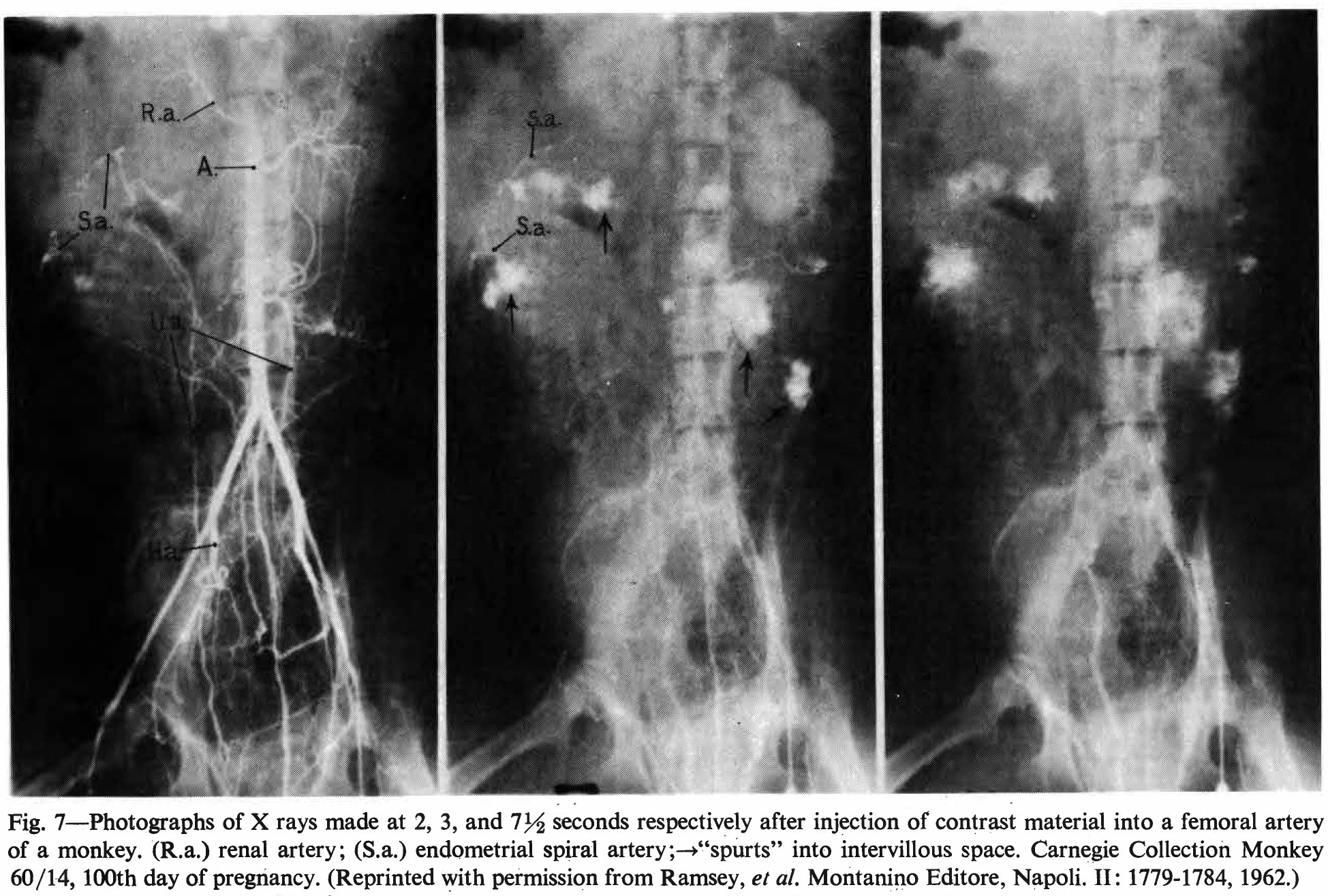 | 1,949 × 1,315 (620 KB) | Z8600021 (talk | contribs) |
You cannot overwrite this file.
File usage
The following page uses this file: