File:Pseudoglandular epithelium.gif
Original file (609 × 635 pixels, file size: 135 KB, MIME type: image/gif)
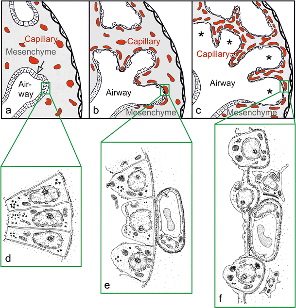
Epithelial structures of lung parenchyma in pseudoglandular, canalicular and saccular stages of lung development
This diagram is a clear demonstration of the key differences in structure of the pulmonary epithelium in the pseudoglandular, canalicular and saccular stages of lung development. In the pseudoglandular stage, it shows bronchial branching into the surrounding mesoderm which is important for later differentiation into specialised alveolar epithelium.
Original legend text
Morphological development of the lung parenchyma during the pseudoglandular, canalicular and saccular stage. The epithelial tubules branch repeatedly during the pseudoglandular stage and penetrate into the surrounding mesenchyme (a, open arrow, branching point). The mesenchyme contains a loose capillary network (a). The epithelium itself is tall and columnar (d). The canalicular stage (b) is characterized (1) by a differentiation of the epithelial cells into type I and type II epithelial cells (e , f), (2) by a widening of the future airways (b), (3) by a multiplication of the capillaries and their first close contacts to the epithelium (b) and (4) by the formation of first future air–blood barriers (e→f). During the saccular stage (c), thick immature inter-airspace septa are formed due to a further condensation of the mesenchyme. The immature septa contain a double-layered capillary network, one layer on either side of the septum. The terminal ends of the bronchial tree represent wide spaces and are called saccules (asterisks). (a–c modified from Caduff et al. 1986; d, e modified from Burri and Weibel 1977, f from Woods and Schittny 2016, by courtesy of Cambridge University Press, New York) [1]
Original source
a-c modified from: Caduff JH, Fischer LC, Burri PH (1986) Scanning electron microscope study of the developing microvasculature in the postnatal rat lung. Anat Rec 216:154–164 d,e modified from: Burri PH, Weibel ER (1977) Ultrastructure and morphometry of the developing lung. In: Hodson WA (ed) Development of the lung. Lung biology in health and disease. M. Dekker, New York, pp 215–268 f from: Woods JC, Schittny JC (2016) Lung structure at preterm and term birth. In: Jobe AH, Whitsett JA, Abman SH (eds) Fetal lung development - clinical correlates & future technologies. Cambridge University Press, New York, pp 126–140
Copyright
This article is distributed under the terms of the Creative Commons Attribution 4.0 International License (http://creativecommons.org/licenses/by/4.0/), which permits unrestricted use, distribution, and reproduction in any medium, provided you give appropriate credit to the original author(s) and the source, provide a link to the Creative Commons license, and indicate if changes were made.
Reference
- ↑ Schittny, J. C. (2017). Development of the lung. Cell and Tissue Research, 367(3), 427–444. http://doi.org/10.1007/s00441-016-2545-0
File history
Click on a date/time to view the file as it appeared at that time.
| Date/Time | Thumbnail | Dimensions | User | Comment | |
|---|---|---|---|---|---|
| current | 10:29, 5 October 2017 | | 609 × 635 (135 KB) | Z5059373 (talk | contribs) |
You cannot overwrite this file.
File usage
The following 2 pages use this file: