File:Progressive myofiber replacement by fibrotic and fat tissue in Dmdmdx rats..jpg
Original file (798 × 606 pixels, file size: 207 KB, MIME type: image/jpeg)
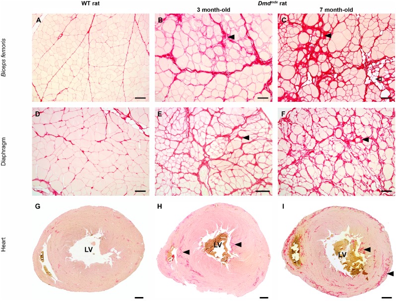
A picrosirius red staining specific for fibrosis was performed on biceps femoris (A–C), respiratory (D–F) and heart muscle (G–I) samples obtained from wild-type littermate controls (WT) and 3 month-old and 7 month-old Dmdmdx rats. Compared to control rats (left panel), a progressive increase in the amount of fibrotic tissue (black arrowhead) was noticed in 3 (mid panel) and 7 month-old Dmdmdx rats (right panel). Note the focal presence of fat tissue infiltration (open arrowhead). In the heart, fibrosis was most marked in papillary muscle of the left ventricle (LV), in the septum and in the ventricular subepicardic area. Picrosirius red staining. Bar = 100 µm (A–F) and 1 mm (G–I).
Copyright: © 2014 Larcher et al. This is an open-access article distributed under the terms of the Creative Commons Attribution License, which permits unrestricted use, distribution, and reproduction in any medium, provided the original author and source are credited.
File history
Click on a date/time to view the file as it appeared at that time.
| Date/Time | Thumbnail | Dimensions | User | Comment | |
|---|---|---|---|---|---|
| current | 22:13, 22 October 2014 | | 798 × 606 (207 KB) | Z3418779 (talk | contribs) | A picrosirius red staining specific for fibrosis was performed on biceps femoris (A–C), respiratory (D–F) and heart muscle (G–I) samples obtained from wild-type littermate controls (WT) and 3 month-old and 7 month-old Dmdmdx rats. Compared to con... |
You cannot overwrite this file.
File usage
The following 3 pages use this file: