File:Process of amniocentesis.jpeg
Original file (3,648 × 2,432 pixels, file size: 2.14 MB, MIME type: image/jpeg)
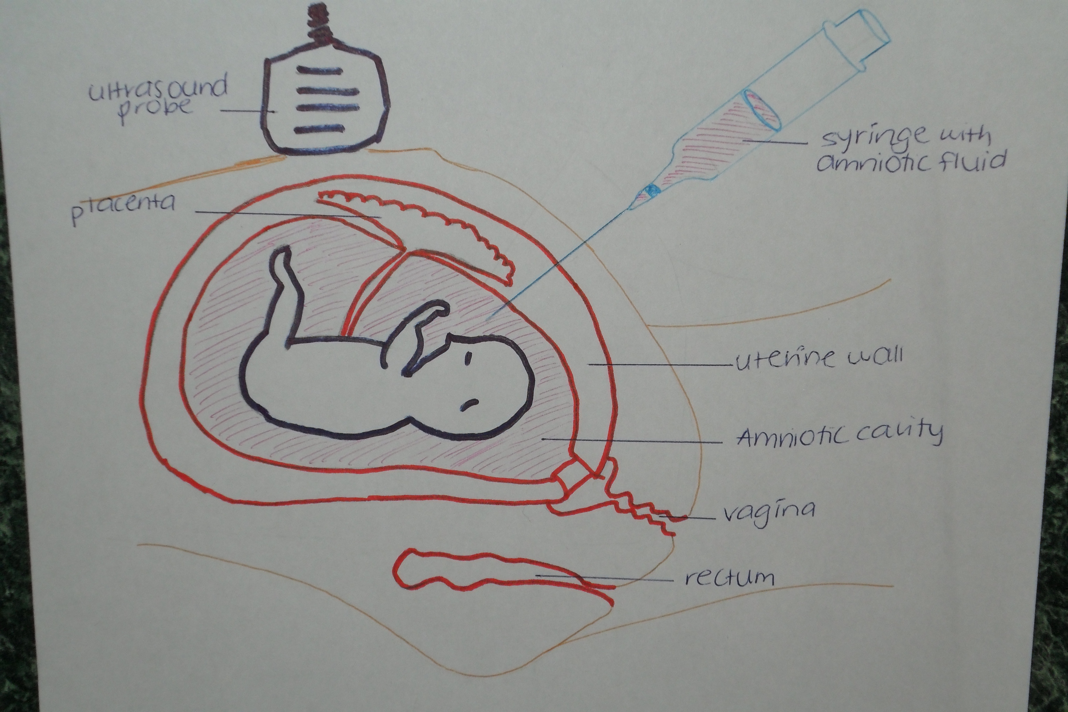
The process of amniocentesis showing the insertion of the needle into the womb to collect amniotic fluid, as well as the use of ultrasound to guide it and prevent harm to the fetus.
Illustration by z3292208.
"Beginning six months after publication, I (student number) grant the public the non-exclusive right to copy, distribute, or display the Work under a Creative Commons Attribution-Noncommercial-Share Alike 3.0 Unported license, as described at http://creativecommons.org/licenses/by-nc-sa/3.0/ and http://creativecommons.org/licenses/by-nc-sa/3.0/legalcode."
File history
Click on a date/time to view the file as it appeared at that time.
| Date/Time | Thumbnail | Dimensions | User | Comment | |
|---|---|---|---|---|---|
| current | 19:22, 31 August 2010 | | 3,648 × 2,432 (2.14 MB) | Z3292208 (talk | contribs) | The process of amniocentesis showing the insertion of the needle into the womb to collect amniotic fluid, as well as the use of ultrasound to guide it and prevent harm to the fetus. Illustration by z3292208. "Beginning six months after publication, I (s |
You cannot overwrite this file.
File usage
The following 2 pages use this file: