File:Primitive streak cell migration.jpg: Difference between revisions
No edit summary |
No edit summary |
||
| Line 3: | Line 3: | ||
(A, B, C) Long-term time-lapse imaging of embryos labelled in Hensen's node (A), the anterior (B) or posterior primitive streak (C) with DiI. Embryos were imaged for 20 hours and still pictures are shown at intervals of 2.5 hours. (D, E, F) Transverse sections of embryos labelled in Hensen's node (D), anterior (E) or posterior streak (F). (G) Summary of cell fates. Red represents Hensen's node and notochord (nc), green represents anterior primitive streak cells and paraxial mesoderm (pm) and blue represents posterior primitive streak cells and lateral plate mesoderm (lpm). n, node; nc, notochord; pm, paraxial mesoderm; s, somites. In (A) asterisks indicate the position of the node which regresses during axis extension. In (A, B, C) arrows show the most recently formed somite and arrowheads in (B, C) indicate the position of the most anterior DiI-labelled cells. | (A, B, C) Long-term time-lapse imaging of embryos labelled in Hensen's node (A), the anterior (B) or posterior primitive streak (C) with DiI. Embryos were imaged for 20 hours and still pictures are shown at intervals of 2.5 hours. (D, E, F) Transverse sections of embryos labelled in Hensen's node (D), anterior (E) or posterior streak (F). (G) Summary of cell fates. Red represents Hensen's node and notochord (nc), green represents anterior primitive streak cells and paraxial mesoderm (pm) and blue represents posterior primitive streak cells and lateral plate mesoderm (lpm). n, node; nc, notochord; pm, paraxial mesoderm; s, somites. In (A) asterisks indicate the position of the node which regresses during axis extension. In (A, B, C) arrows show the most recently formed somite and arrowheads in (B, C) indicate the position of the most anterior DiI-labelled cells. | ||
Original file name: 1471-213X-8-63-1.jpg | :Original file name: 1471-213X-8-63-1.jpg | ||
The migration of paraxial and lateral plate mesoderm cells emerging from the late primitive streak is controlled by different Wnt signals. Sweetman D, Wagstaff L, Cooper O, Weijer C, Münsterberg A. BMC Dev Biol. 2008 Jun 9;8:63. [http://www.ncbi.nlm.nih.gov/pubmed/18541012 PMID: 18541012] | |||
'''Reference:''' The migration of paraxial and lateral plate mesoderm cells emerging from the late primitive streak is controlled by different Wnt signals. Sweetman D, Wagstaff L, Cooper O, Weijer C, Münsterberg A. BMC Dev Biol. 2008 Jun 9;8:63. [http://www.ncbi.nlm.nih.gov/pubmed/18541012 PMID: 18541012] | |||
:"We suggest that the distinct behaviours of paraxial and lateral mesoderm precursors are regulated by the opposing actions of Wnt5a and Wnt3a as they leave the primitive streak in neurula stage embryos. Our data suggests that Wnt5a acts via prickle to cause migration of cells from the posterior streak. In the anterior streak, this is antagonised by Wnt3a to generate non-migratory medial mesoderm." | :"We suggest that the distinct behaviours of paraxial and lateral mesoderm precursors are regulated by the opposing actions of Wnt5a and Wnt3a as they leave the primitive streak in neurula stage embryos. Our data suggests that Wnt5a acts via prickle to cause migration of cells from the posterior streak. In the anterior streak, this is antagonised by Wnt3a to generate non-migratory medial mesoderm." | ||
This is an Open Access article distributed under the terms of the Creative Commons Attribution License (http://creativecommons.org/licenses/by/2.0), which permits unrestricted use, distribution, and reproduction in any medium, provided the original work is properly cited. | This is an Open Access article distributed under the terms of the Creative Commons Attribution License (http://creativecommons.org/licenses/by/2.0), which permits unrestricted use, distribution, and reproduction in any medium, provided the original work is properly cited. | ||
Revision as of 20:41, 3 August 2009
DiI labelling in HH8 embryos reveals behaviour and trajectories of cells from the primitive streak.
(A, B, C) Long-term time-lapse imaging of embryos labelled in Hensen's node (A), the anterior (B) or posterior primitive streak (C) with DiI. Embryos were imaged for 20 hours and still pictures are shown at intervals of 2.5 hours. (D, E, F) Transverse sections of embryos labelled in Hensen's node (D), anterior (E) or posterior streak (F). (G) Summary of cell fates. Red represents Hensen's node and notochord (nc), green represents anterior primitive streak cells and paraxial mesoderm (pm) and blue represents posterior primitive streak cells and lateral plate mesoderm (lpm). n, node; nc, notochord; pm, paraxial mesoderm; s, somites. In (A) asterisks indicate the position of the node which regresses during axis extension. In (A, B, C) arrows show the most recently formed somite and arrowheads in (B, C) indicate the position of the most anterior DiI-labelled cells.
- Original file name: 1471-213X-8-63-1.jpg
Reference: The migration of paraxial and lateral plate mesoderm cells emerging from the late primitive streak is controlled by different Wnt signals. Sweetman D, Wagstaff L, Cooper O, Weijer C, Münsterberg A. BMC Dev Biol. 2008 Jun 9;8:63. PMID: 18541012
- "We suggest that the distinct behaviours of paraxial and lateral mesoderm precursors are regulated by the opposing actions of Wnt5a and Wnt3a as they leave the primitive streak in neurula stage embryos. Our data suggests that Wnt5a acts via prickle to cause migration of cells from the posterior streak. In the anterior streak, this is antagonised by Wnt3a to generate non-migratory medial mesoderm."
This is an Open Access article distributed under the terms of the Creative Commons Attribution License (http://creativecommons.org/licenses/by/2.0), which permits unrestricted use, distribution, and reproduction in any medium, provided the original work is properly cited.
BMC Dev Biol. 2008; 8: 63. Published online 2008 June 9. doi: 10.1186/1471-213X-8-63. Copyright © 2008 Sweetman et al; licensee BioMed Central Ltd.
File history
Click on a date/time to view the file as it appeared at that time.
| Date/Time | Thumbnail | Dimensions | User | Comment | |
|---|---|---|---|---|---|
| current | 20:38, 3 August 2009 | 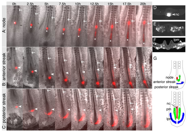 | 600 × 420 (92 KB) | S8600021 (talk | contribs) | DiI labelling in HH8 embryos reveals behaviour and trajectories of cells from the primitive streak. (A, B, C) Long-term time-lapse imaging of embryos labelled in Hensen's node (A), the anterior (B) or posterior primitive streak (C) with DiI. Embryos wer |
You cannot overwrite this file.
File usage
The following page uses this file: