File:Primary Brain.png
From Embryology
Size of this preview: 800 × 521 pixels. Other resolution: 1,005 × 655 pixels.
Original file (1,005 × 655 pixels, file size: 681 KB, MIME type: image/png)
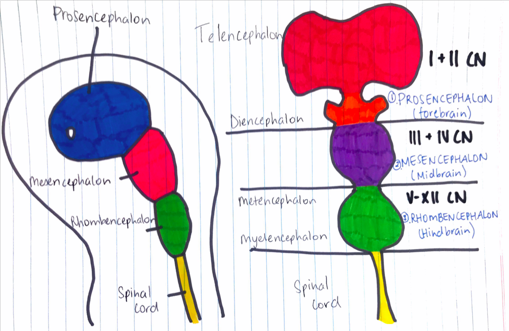
Primary Brain Vesicles
In week 4, 3 primary brain vesicles are formed; forebrain (prosencephalon) midbrain (mesencephalon), and hindbrain (rhombencephalon). These structures then differentiate into 5 secondary brain vesicles during week 5.[1]
Copyright & Reference
Drawn by Student z5076158, however base on: </reference>
- Note - This image was originally uploaded as part of an undergraduate science student project and may contain inaccuracies in either description or acknowledgements. Students have been advised in writing concerning the reuse of content and may accidentally have misunderstood the original terms of use. If image reuse on this non-commercial educational site infringes your existing copyright, please contact the site editor for immediate removal.
- ↑ Khanal, L. 2017. Development of nervous system. Retrieved October 20 from, https://www.slideshare.net/NamXal1/development-of-nervous-system
File history
Click on a date/time to view the file as it appeared at that time.
| Date/Time | Thumbnail | Dimensions | User | Comment | |
|---|---|---|---|---|---|
| current | 09:54, 25 October 2017 | | 1,005 × 655 (681 KB) | Z5076158 (talk | contribs) | In week 4, 3 primary brain vesicles are formed; forebrain (prosencephalon) midbrain (mesencephalon), and hindbrain (rhombencephalon). These structures then differentiate into 5 secondary brain vesicles during week 5. Drawn by Student z5076158 {{Templ... |
You cannot overwrite this file.
File usage
The following 2 pages use this file: