File:Prentiss 286.jpg: Difference between revisions
No edit summary |
|||
| Line 3: | Line 3: | ||
(Allea Thomson) | (Allea Thomson) | ||
Diagrammatic outline of the organs of circulation in the fetus of ail months (Allen Thomson). RA, Right atrium of the heart; RV, right ventricle; LA, left atrium; Ev, valve of inf. vena cava; LV, left ventricle; L, liver; K, left kidney; I, small intestine; a, arch of the aorta; a', its dorsal part; a", lower end; | Diagrammatic outline of the organs of circulation in the fetus of ail months (Allen Thomson). RA, Right atrium of the heart; RV, right ventricle; LA, left atrium; Ev, valve of inf. vena cava; LV, left ventricle; L, liver; K, left kidney; I, small intestine; a, arch of the aorta; a', its dorsal part; a", lower end; vcs, superior vena cava; vci, inferior vena cava where it joins the right atrium; vet' its lower end; i, subclavian vessels; j, right jugular vein;c, common carotid arteries; four tnirvcd dotted arrow lines are carried through the aortic and pulmonary opening and ihe atrio-ventricular orifices; da, opposite to the one passing through the pulmonary artery, marks the place of the ductus arteriosus; a similar arrow line is shown passing from the vena cava inferior through the fossa ovalis of the right atrium and the foramen ovale into the left atrium; hv, the hepatic veins; vp, vena ports; i to vci, the ductus venosus; uv, umbilical vein; ita, umbilical arteries; ur, umbilical cord cut short; i, i', iliac vessels. | ||
Revision as of 02:58, 28 September 2012
Fig. 286 Diagrammatic outline of the organs of circulation in the fetus of six months
(Allea Thomson)
Diagrammatic outline of the organs of circulation in the fetus of ail months (Allen Thomson). RA, Right atrium of the heart; RV, right ventricle; LA, left atrium; Ev, valve of inf. vena cava; LV, left ventricle; L, liver; K, left kidney; I, small intestine; a, arch of the aorta; a', its dorsal part; a", lower end; vcs, superior vena cava; vci, inferior vena cava where it joins the right atrium; vet' its lower end; i, subclavian vessels; j, right jugular vein;c, common carotid arteries; four tnirvcd dotted arrow lines are carried through the aortic and pulmonary opening and ihe atrio-ventricular orifices; da, opposite to the one passing through the pulmonary artery, marks the place of the ductus arteriosus; a similar arrow line is shown passing from the vena cava inferior through the fossa ovalis of the right atrium and the foramen ovale into the left atrium; hv, the hepatic veins; vp, vena ports; i to vci, the ductus venosus; uv, umbilical vein; ita, umbilical arteries; ur, umbilical cord cut short; i, i', iliac vessels.
In this diagram the arteries are conventionally colored red and the veins blue, but these colors are not intended to indicate the nature of the blood conveyed by the reflective vessels.
| Embryology - 16 Apr 2024 |
|---|
| Google Translate - select your language from the list shown below (this will open a new external page) |
|
العربية | català | 中文 | 中國傳統的 | français | Deutsche | עִברִית | हिंदी | bahasa Indonesia | italiano | 日本語 | 한국어 | မြန်မာ | Pilipino | Polskie | português | ਪੰਜਾਬੀ ਦੇ | Română | русский | Español | Swahili | Svensk | ไทย | Türkçe | اردو | ייִדיש | Tiếng Việt These external translations are automated and may not be accurate. (More? About Translations) |
Prentiss CW. and Arey LB. A laboratory manual and text-book of embryology. (1918) W.B. Saunders Company, Philadelphia and London.
| Historic Disclaimer - information about historic embryology pages |
|---|
 |
- (Text at early stage of editing, images to be added)
| Historic Disclaimer - information about historic embryology pages |
|---|
|
File history
Click on a date/time to view the file as it appeared at that time.
| Date/Time | Thumbnail | Dimensions | User | Comment | |
|---|---|---|---|---|---|
| current | 16:37, 13 September 2012 | 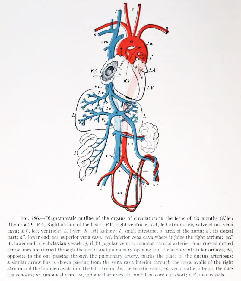 | 799 × 933 (121 KB) | Z8600021 (talk | contribs) |
You cannot overwrite this file.
File usage
The following page uses this file: