File:Polar Body Biopsy.jpeg
Polar_Body_Biopsy.jpeg (500 × 375 pixels, file size: 74 KB, MIME type: image/jpeg)
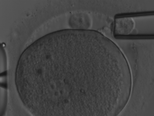
Polar Body Biopsy
The presence of a faint but clearly identifiable strand connecting PB2 to the oolemma. The biopsy was performed ∼9 h after ICSI.
(Original figure legend, image based on data from Magli et al. PMID 21908464 )
Reference
<pubmed>21908464</pubmed>| [Polar body array CGH for prediction of the status of the corresponding oocyte. Part II: technical aspects]
Copyright
© The Author 2011. Published by Oxford University Press on behalf of the European Society of Human Reproduction and Embryology.
This is an Open Access article distributed under the terms of the Creative Commons Attribution Non-Commercial License (http://creativecommons.org/licenses/by-nc/2.5), which permits unrestricted non-commercial use, distribution, and reproduction in any medium, provided the original work is properly cited
Figure 1: Der29501.jpg
- Note - This image was originally uploaded as part of an undergraduate science student project and may contain inaccuracies in either description or acknowledgements. Students have been advised in writing concerning the reuse of content and may accidentally have misunderstood the original terms of use. If image reuse on this non-commercial educational site infringes your existing copyright, please contact the site editor for immediate removal.
File history
Click on a date/time to view the file as it appeared at that time.
| Date/Time | Thumbnail | Dimensions | User | Comment | |
|---|---|---|---|---|---|
| current | 07:00, 1 October 2015 | | 500 × 375 (74 KB) | Z5088434 (talk | contribs) | PMID 21908464 Der29501.jpg |
You cannot overwrite this file.
File usage
The following 2 pages use this file: