File:Placental villi 2.jpg
Original file (1,280 × 1,024 pixels, file size: 70 KB, MIME type: image/jpeg)
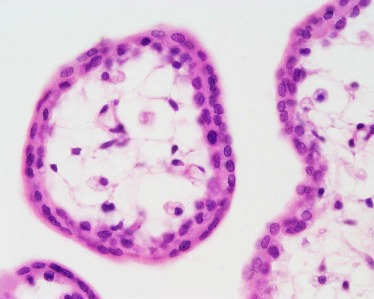
Human Placental Villi First Trimester
- cross-section image.
- Villi are cover in shell of cytotrophoblast cells.
- Villi core contains mainly mesenchyme cells.
- Links: Image - First trimester villi overview | Image - First trimester villi detail | | Image - Term villi detail | Fetal Development | Placenta Development
placenta, first trimester, human H&E
reproductive system, female, chorionic villi, cytotrophoblast, syncytiotrophoblast
original file name Ple41he.jpg
Links: Histology | Histology Stains | Blue Histology images copyright Lutz Slomianka 1998-2009. The literary and artistic works on the original Blue Histology website may be reproduced, adapted, published and distributed for non-commercial purposes. See also the page Histology Stains.
Cite this page: Hill, M.A. (2024, April 19) Embryology Placental villi 2.jpg. Retrieved from https://embryology.med.unsw.edu.au/embryology/index.php/File:Placental_villi_2.jpg
- © Dr Mark Hill 2024, UNSW Embryology ISBN: 978 0 7334 2609 4 - UNSW CRICOS Provider Code No. 00098G
File history
Click on a date/time to view the file as it appeared at that time.
| Date/Time | Thumbnail | Dimensions | User | Comment | |
|---|---|---|---|---|---|
| current | 02:36, 1 April 2012 | | 1,280 × 1,024 (70 KB) | Z8600021 (talk | contribs) | |
| 16:34, 3 August 2009 | 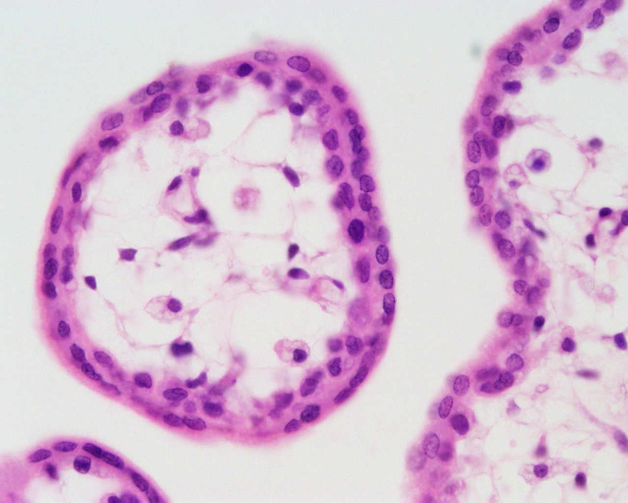 | 1,280 × 1,024 (230 KB) | MarkHill (talk | contribs) | Human placental villi cross-section placenta, first trimester, human H&E reproductive system, female, chorionic villi, cytotrophoblast, syncytiotrophoblast original file name Ple41he.jpg Image Source: UWA Blue Histology http://www.lab.anhb.uwa.edu.au/ |
You cannot overwrite this file.
File usage
The following 4 pages use this file: