File:Placental membranes.jpg: Difference between revisions
From Embryology
| Line 5: | Line 5: | ||
* '''amniotic sac''' - formed by the amniotic membrane (ectoderm and extra-embryonic mesoderm) completely surrounding the surrounding the embryo. | * '''amniotic sac''' - formed by the amniotic membrane (ectoderm and extra-embryonic mesoderm) completely surrounding the surrounding the embryo. | ||
* '''yolk sac''' - the yolk membrane (endoderm and extra-embryonic mesoderm) attached to the embryo at the umbilicus and continuous with the midgut. | * '''yolk sac''' - the yolk membrane (endoderm and extra-embryonic mesoderm) attached to the embryo at the umbilicus and continuous with the midgut. | ||
* '''chorionic cavity''' - membrane (extra-embryonic mesoderm) has been removed but is represented by the black space outside the amniotic and yolk sac and part of the remaining chorionic villi region. | * '''chorionic cavity''' - membrane (extra-embryonic mesoderm) has been removed but is represented by the black space outside the amniotic and yolk sac and part of the remaining chorionic villi region, labeled with placental vessels. | ||
Revision as of 07:46, 15 May 2014
Embryo with Placental Membranes
By the external appearance of the embryo appears to be about Week 7 (GA week 9) either Carnegie stage 18 or Carnegie stage 19
- amniotic sac - formed by the amniotic membrane (ectoderm and extra-embryonic mesoderm) completely surrounding the surrounding the embryo.
- yolk sac - the yolk membrane (endoderm and extra-embryonic mesoderm) attached to the embryo at the umbilicus and continuous with the midgut.
- chorionic cavity - membrane (extra-embryonic mesoderm) has been removed but is represented by the black space outside the amniotic and yolk sac and part of the remaining chorionic villi region, labeled with placental vessels.
Image Source: UNSW Embryology, no reproduction without permission.
- Carnegie Stages: 1 | 2 | 3 | 4 | 5 | 6 | 7 | 8 | 9 | 10 | 11 | 12 | 13 | 14 | 15 | 16 | 17 | 18 | 19 | 20 | 21 | 22 | 23 | About Stages | Timeline
Cite this page: Hill, M.A. (2024, April 24) Embryology Placental membranes.jpg. Retrieved from https://embryology.med.unsw.edu.au/embryology/index.php/File:Placental_membranes.jpg
- © Dr Mark Hill 2024, UNSW Embryology ISBN: 978 0 7334 2609 4 - UNSW CRICOS Provider Code No. 00098G
File history
Click on a date/time to view the file as it appeared at that time.
| Date/Time | Thumbnail | Dimensions | User | Comment | |
|---|---|---|---|---|---|
| current | 23:36, 16 August 2009 | 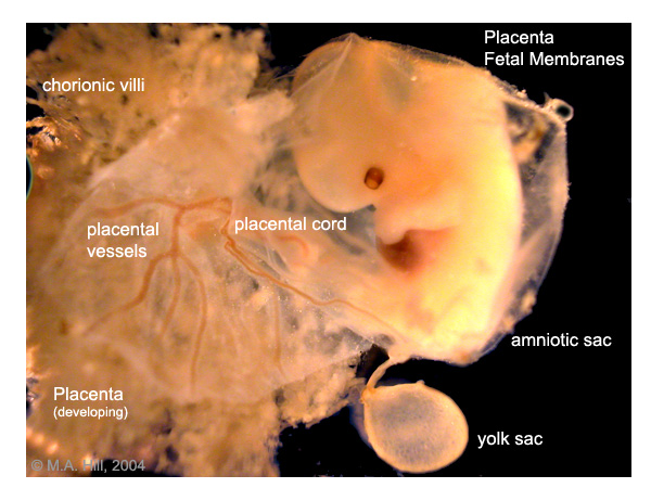 | 600 × 450 (99 KB) | S8600021 (talk | contribs) | Image Source: UNSW Embryology, no reproduction without permission. PlMembraneW600.jpg http://embryology.med.unsw.edu.au/Notes/images/placenta/plMembraneW600.jpg |
You cannot overwrite this file.
File usage
The following 16 pages use this file:
- 2009 Lecture 8
- 2010 BGD Lecture - Development of the Embryo/Fetus 1
- 2010 Group Project 3
- 2010 Lecture 8
- ANAT2341 Lab 4 - Implantation and Villi Development
- ASA Meeting 2013 - Placenta
- BGDA Lecture - Development of the Embryo/Fetus 1
- BGDA Practical 3 - Extraembryonic Spaces
- BGDA Practical Placenta - Villi Development
- Foundations Practical - Week 1 to 8
- Human System Development
- Lecture - Early Vascular Development
- P
- Placenta - Membranes
- Placenta Development
- Yolk Sac Development