File:Placenta anchoring villi.jpg: Difference between revisions
No edit summary |
|||
| Line 12: | Line 12: | ||
:'''Links:''' [[Implantation]] | [[Placenta Development]] | :'''Links:''' [[Implantation]] | [[Placenta Development]] | ||
Image Source: UNSW Embryology, no reproduction without permission. | |||
Image Source: UNSW Embryology, no reproduction without permission. | |||
[[Category:Placenta]] [[Category:Histology]] | [[Category:Placenta]] [[Category:Histology]] | ||
Revision as of 13:56, 9 May 2011
Placenta Anchoring Villi
Histological image showing the junctional region between the trophoblast shell of the conceptus and the maternal decidua.
In week 2, the trophoblast shell cells proliferate and form a syncitiotrophoblast and cytotrophoblast layer around he conceptus. Syncitiotrophoblast cells migrate into the uterine wall, forming maternal blood-filled spaces (lacunae).
Placentation begins once the conceptus begins to implant in the uterine wall and the placenta will have both a fetal and a maternal component. The fetal component begins as villi form. The fetal chorion will form two regions: smooth chorion (chorion laeve) and villous chorion (chorion frondosum).
The maternal component is formed by the decidualization of the endometrium.
- Links: Implantation | Placenta Development
Image Source: UNSW Embryology, no reproduction without permission.
File history
Click on a date/time to view the file as it appeared at that time.
| Date/Time | Thumbnail | Dimensions | User | Comment | |
|---|---|---|---|---|---|
| current | 14:37, 3 August 2009 | 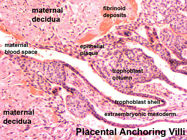 | 600 × 450 (167 KB) | MarkHill (talk | contribs) | Placenta anchoring villi Histological image showing the junctional region between the trophoblast shell of the conceptus and the maternal decidua. In week 2, the trophoblast shell cells proliferate and form a syncitiotrophoblast and cytotrophoblast lay |
You cannot overwrite this file.
File usage
The following 31 pages use this file:
- 2009 Lecture 4
- 2009 Lecture 8
- 2010 BGD Lecture - Development of the Embryo/Fetus 1
- 2010 Lab 4
- 2010 Lecture 4
- 2010 Lecture 8
- 2011 Lab 4
- A
- ACPS Seminar 2014 - Implantation
- ANAT2341 Lab 4 - Decidua and Cord
- ANAT2341 Lab 4 - Implantation and Villi Development
- ASA Meeting 2013 - Placenta
- BGDA Lecture - Development of the Embryo/Fetus 1
- BGDA Practical Placenta - Maternal Decidua
- BGDA Practical Placenta - Villi Development
- D
- F
- Implantation
- Lecture - Placenta Development
- Lecture - Week 1 and 2 Development
- Lecture - Week 3 Development
- Placenta - Histology
- Placenta - Maternal Decidua
- Placenta - Membranes
- Placenta Development
- Trophoblast - Protein Expression
- Week 2
- Yolk Sac Development
- Talk:ANAT2341 Lab 4 - Implantation and Villi Development
- Talk:Lecture - Week 3 Development
- User:Z5014754