File:Peyer's patch 02.jpg: Difference between revisions
From Embryology
No edit summary |
m (→Peyer's Patch) |
||
| (8 intermediate revisions by the same user not shown) | |||
| Line 1: | Line 1: | ||
==Peyer's Patch== | ==Peyer's Patch== | ||
{| | |||
| width=40%| | |||
* '''IELs''' - intraepithelial lymphocytes | |||
** antigen-experienced T cells (not requiring to be primed). | |||
** balance protective immunity with an ability to safeguard the integrity of the epithelial barrier<ref><pubmed>21681197</pubmed>| [http://www.ncbi.nlm.nih.gov/pmc/articles/PMC3140792 Nat Rev Immunol.]</ref> | |||
** contact with antigen immediately release cytokines. | |||
** can kill infected target cells. | |||
| width=40%| | |||
* '''M-cell''' - microfold cell | |||
** found in the follicle-associated epithelium of the Peyer's patch. | |||
** function to transport gut lumen organisms and particles to immune cells across the epithelial barrier. | |||
|} | |||
{{Immune Images 2}} | |||
<references/> | |||
{{Blue Histology}} | |||
[[Category:Immune]] [[Category:Gastrointestinal Tract]] | [[Category:Immune]] [[Category:Gastrointestinal Tract]] | ||
Latest revision as of 12:14, 26 January 2015
Peyer's Patch
|
|
- Immune Images: Oesophagus MALT | Colon MALT | Peyer's patch overview | Peyer's patch detail | Cartoon - IEL development | Cartoon - IEL function | Cartoon - IEL differentiation | Mesenteric Lymph Nodes overview | Palatine Tonsil | Tonsil | Immune System Development
- ↑ <pubmed>21681197</pubmed>| Nat Rev Immunol.
Links: Histology | Histology Stains | Blue Histology images copyright Lutz Slomianka 1998-2009. The literary and artistic works on the original Blue Histology website may be reproduced, adapted, published and distributed for non-commercial purposes. See also the page Histology Stains.
Cite this page: Hill, M.A. (2024, April 17) Embryology Peyer's patch 02.jpg. Retrieved from https://embryology.med.unsw.edu.au/embryology/index.php/File:Peyer%27s_patch_02.jpg
- © Dr Mark Hill 2024, UNSW Embryology ISBN: 978 0 7334 2609 4 - UNSW CRICOS Provider Code No. 00098G
File history
Click on a date/time to view the file as it appeared at that time.
| Date/Time | Thumbnail | Dimensions | User | Comment | |
|---|---|---|---|---|---|
| current | 10:05, 24 February 2012 | 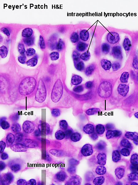 | 450 × 600 (69 KB) | Z8600021 (talk | contribs) | |
| 08:23, 24 December 2010 |  | 300 × 400 (41 KB) | S8600021 (talk | contribs) | Peyer's patch 01.jpg Pey102he.jpg |
You cannot overwrite this file.
File usage
The following 3 pages use this file: