File:Pericardial Development 4-6 Gestation weeks .jpg

Original file (727 × 834 pixels, file size: 102 KB, MIME type: image/jpeg)
PMCID: PMC4374196
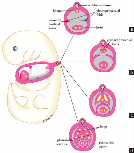
Pericardial Development
Illustration of normal pericardial development from the fourth to sixth week of gestation. (a) At 4 weeks of gestation, the laterally positioned pleuropericardial folds are developed. (b) During the 5th week of gestation, the pleuropericardial folds grow toward the midline while the root of each fold migrates ventrally. (c) At the end of the 5th week, the pleuropericardial folds fuse, partitioning the thoracic cavity into a pericardial cavity and two partially formed pleural cavities. Note that union of the pleuropericardial folds and the root of the lungs also occurs during this time. (d) The lungs continue to extend anteriorly to the front of the heart.
Reference
<PMCID>4374196</PMCID>
Copyright : © 2015 Koo CW.
This is an open-access article distributed under the terms of the Creative Commons Attribution License, which permits unrestricted use, distribution, and reproduction in any medium, provided the original author and source are credited.
File history
Click on a date/time to view the file as it appeared at that time.
| Date/Time | Thumbnail | Dimensions | User | Comment | |
|---|---|---|---|---|---|
| current | 14:45, 26 August 2017 | | 727 × 834 (102 KB) | Z5076019 (talk | contribs) | PMCID: PMC4374196 |
You cannot overwrite this file.
File usage
The following 2 pages use this file: