File:Patten051.jpg: Difference between revisions
({{Template:Patten_1920_Figures}}) |
No edit summary |
||
| Line 1: | Line 1: | ||
==Fig. 51. Schematic diagrams to show the relations of pronephros, mesonephros, and metanephros at various stages of development== | |||
In the first stage represented (Fig. 51, A) only the pronephros has been established. It consists of a group of tubules emptying into a common duct, called the pronephric duct. The pronephric ducts of either side are formed first at the level of the pronephric tubules and then extend caudad, eventually reaching and opening into the cloaca (See arrows in Fig. 51, A). | |||
As the pronephric ducts are extended caudal to the level at which pronephric tubules are formed they come in close proximity to the developing mesonephric tubules. In their growth the mesonephric tubules extend toward the pronephric ducts and soon open into them (Fig. 51, B). Meanwhile the pronephric tubules begin to degenerate. Thus the ducts which originally arose in connection with the pronephros are appropriated by the developing mesonephros. After the degeneration of the pronephric tubules these same ducts are called the mesonephric ducts because of their new associations (Fig. 51 , C). | |||
At a considerably later stage outgrowths develop from the mesonephric ducts near their cloacal ends (Fig. 51, C). These outgrowths form the ducts of the metanephroi. They grow cephalo-laterad and eventually connect with the third group of tubules developed from the intermediate mesoderm, the metanephric tubules (Fig. 51, D). With the establishment of the metanephroi or permanent kidneys the mesonephroi begin to degenerate. The only parts of the mesonephric system to persist, except in vestigial form, are some of the ducts and tubules which in the male are appropriated by the testis as a duct system. | |||
{{Template:Patten_1920_Figures}} | {{Template:Patten_1920_Figures}} | ||
[[Category:Renal]] | |||
Latest revision as of 08:23, 29 July 2011
Fig. 51. Schematic diagrams to show the relations of pronephros, mesonephros, and metanephros at various stages of development
In the first stage represented (Fig. 51, A) only the pronephros has been established. It consists of a group of tubules emptying into a common duct, called the pronephric duct. The pronephric ducts of either side are formed first at the level of the pronephric tubules and then extend caudad, eventually reaching and opening into the cloaca (See arrows in Fig. 51, A).
As the pronephric ducts are extended caudal to the level at which pronephric tubules are formed they come in close proximity to the developing mesonephric tubules. In their growth the mesonephric tubules extend toward the pronephric ducts and soon open into them (Fig. 51, B). Meanwhile the pronephric tubules begin to degenerate. Thus the ducts which originally arose in connection with the pronephros are appropriated by the developing mesonephros. After the degeneration of the pronephric tubules these same ducts are called the mesonephric ducts because of their new associations (Fig. 51 , C).
At a considerably later stage outgrowths develop from the mesonephric ducts near their cloacal ends (Fig. 51, C). These outgrowths form the ducts of the metanephroi. They grow cephalo-laterad and eventually connect with the third group of tubules developed from the intermediate mesoderm, the metanephric tubules (Fig. 51, D). With the establishment of the metanephroi or permanent kidneys the mesonephroi begin to degenerate. The only parts of the mesonephric system to persist, except in vestigial form, are some of the ducts and tubules which in the male are appropriated by the testis as a duct system.
- Links: Introduction | Gametes and Fertilization | Segmentation | Entoderm | Primitive Streak and Mesoderm | Primitive Streak to Somites | 24 Hours | 24 to 33 Hours | 33 to 39 Hours | 40 to 50 Hours | Extra-embryonic Membranes | 50 to 55 Hours | Day 3 to 4 | References | All Figures
| Historic Disclaimer - information about historic embryology pages |
|---|
 |
Reference
Patten BM. The Early Embryology of the Chick. (1920) Philadelphia: P. Blakiston's Son and Co.
Cite this page: Hill, M.A. (2024, April 25) Embryology Patten051.jpg. Retrieved from https://embryology.med.unsw.edu.au/embryology/index.php/File:Patten051.jpg
- © Dr Mark Hill 2024, UNSW Embryology ISBN: 978 0 7334 2609 4 - UNSW CRICOS Provider Code No. 00098G
File history
Click on a date/time to view the file as it appeared at that time.
| Date/Time | Thumbnail | Dimensions | User | Comment | |
|---|---|---|---|---|---|
| current | 02:05, 17 January 2011 | 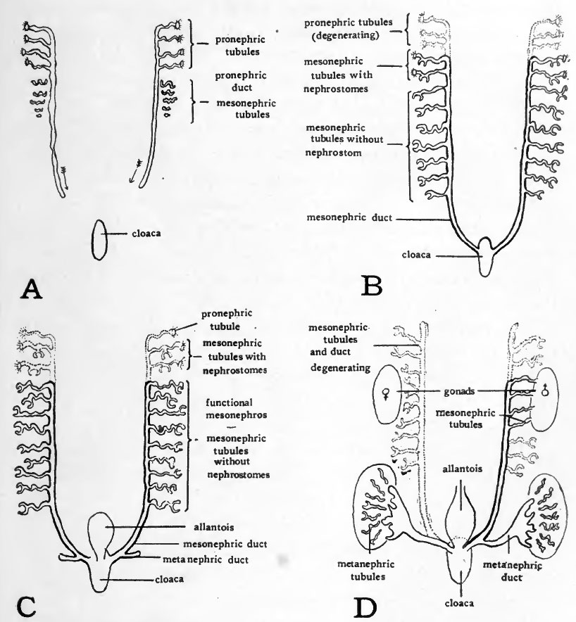 | 818 × 884 (120 KB) | S8600021 (talk | contribs) | {{Template:Patten_1920_Figures}} |
You cannot overwrite this file.
File usage
The following 3 pages use this file: