File:Patten037.jpg: Difference between revisions
({{Template:Patten_1920_Figures}}) |
No edit summary |
||
| Line 1: | Line 1: | ||
==Fig. 37. Drawings from transverse sections to show origin of neural crest cells== | |||
The location of the area drawn is indicated on the small sketch to the left of each drawing. | |||
'''A''' anterior rhombencephalic region of 30-hour chick | |||
'''B''' posterior rhombencephalic region of 36-hour chick | |||
'''C''' mid-dorsal region of cord in 55-hour chick. | |||
The neural crest should not be confused with the margin of the neural fold with which it is associated before the closure of the neural tube. The margin of the neural fold involves cells which go into the superficial ectoderm and into the neural tube, as well as those which are concerned in the formation of the neural crest. | |||
When first established the neural crest is continuous anteroposteriorly. As development proceeds, the cells of the neural crest migrate ventro-laterally on either side of the spinal cord (Fig. 37, C), and at the same time become segmen tally clustered. The segmen tally arranged cell groups thus derived from the neural crest give rise to the dorsal root ganglia of the spinal nerves, and in the head region to the ganglia of the sensory cranial nerves. | |||
(For a later stage of the dorsal root ganglia see Figure 44.) | |||
{{Template:Patten_1920_Figures}} | {{Template:Patten_1920_Figures}} | ||
Latest revision as of 09:33, 29 July 2011
Fig. 37. Drawings from transverse sections to show origin of neural crest cells
The location of the area drawn is indicated on the small sketch to the left of each drawing.
A anterior rhombencephalic region of 30-hour chick
B posterior rhombencephalic region of 36-hour chick
C mid-dorsal region of cord in 55-hour chick.
The neural crest should not be confused with the margin of the neural fold with which it is associated before the closure of the neural tube. The margin of the neural fold involves cells which go into the superficial ectoderm and into the neural tube, as well as those which are concerned in the formation of the neural crest.
When first established the neural crest is continuous anteroposteriorly. As development proceeds, the cells of the neural crest migrate ventro-laterally on either side of the spinal cord (Fig. 37, C), and at the same time become segmen tally clustered. The segmen tally arranged cell groups thus derived from the neural crest give rise to the dorsal root ganglia of the spinal nerves, and in the head region to the ganglia of the sensory cranial nerves.
(For a later stage of the dorsal root ganglia see Figure 44.)
- Links: Introduction | Gametes and Fertilization | Segmentation | Entoderm | Primitive Streak and Mesoderm | Primitive Streak to Somites | 24 Hours | 24 to 33 Hours | 33 to 39 Hours | 40 to 50 Hours | Extra-embryonic Membranes | 50 to 55 Hours | Day 3 to 4 | References | All Figures
| Historic Disclaimer - information about historic embryology pages |
|---|
 |
Reference
Patten BM. The Early Embryology of the Chick. (1920) Philadelphia: P. Blakiston's Son and Co.
Cite this page: Hill, M.A. (2024, April 16) Embryology Patten037.jpg. Retrieved from https://embryology.med.unsw.edu.au/embryology/index.php/File:Patten037.jpg
- © Dr Mark Hill 2024, UNSW Embryology ISBN: 978 0 7334 2609 4 - UNSW CRICOS Provider Code No. 00098G
File history
Click on a date/time to view the file as it appeared at that time.
| Date/Time | Thumbnail | Dimensions | User | Comment | |
|---|---|---|---|---|---|
| current | 02:16, 17 January 2011 | 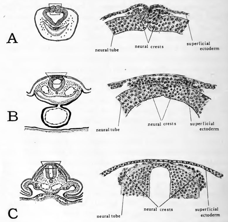 | 778 × 758 (107 KB) | S8600021 (talk | contribs) | {{Template:Patten_1920_Figures}} |
You cannot overwrite this file.
File usage
The following 2 pages use this file: