File:Patten036.jpg: Difference between revisions
({{Template:Patten_1920_Figures}}) |
No edit summary |
||
| Line 1: | Line 1: | ||
==Fig. 36. Diagrams of transverse sections of 55-hour (30-somiite) chick== | |||
The location of the sections is indicated on an outline sketch of the entire embryo. | |||
While the secondary optic vesicles are usually spoken of as the optic cups, they are not complete cups. The invagination which gives rise to the secondary optic vesicles, instead of beginning at the most lateral point in the primary optic vesicles, begins at a point somewhat toward their ventral surface and is directed mesiodorsad. As a result the optic cups are formed without any lip on their ventral aspect. They may be likened to cups with a segment broken out of one side. This gap in the optic cup is the choroid fissure (Fig. 35). In Figure 36, B, a section is shown which passes through the head of the embryo on a slight slant so that the right optic cup, being cut to one side of the choroid fissure appears complete while the left optic cup being cut in the region of the fissure shows no ventral lip. | |||
The infolding process by which the optic cups are formed from the primary optic vesicles is continued to the region of the optic stalks. As a result the optic stalks are infolded so that their ventral surfaces become grooved. Later in development the optic nerves and blood vessels come to lie in the grooves thus formed in the optic stalks. | |||
{{Template:Patten_1920_Figures}} | {{Template:Patten_1920_Figures}} | ||
Latest revision as of 09:31, 29 July 2011
Fig. 36. Diagrams of transverse sections of 55-hour (30-somiite) chick
The location of the sections is indicated on an outline sketch of the entire embryo.
While the secondary optic vesicles are usually spoken of as the optic cups, they are not complete cups. The invagination which gives rise to the secondary optic vesicles, instead of beginning at the most lateral point in the primary optic vesicles, begins at a point somewhat toward their ventral surface and is directed mesiodorsad. As a result the optic cups are formed without any lip on their ventral aspect. They may be likened to cups with a segment broken out of one side. This gap in the optic cup is the choroid fissure (Fig. 35). In Figure 36, B, a section is shown which passes through the head of the embryo on a slight slant so that the right optic cup, being cut to one side of the choroid fissure appears complete while the left optic cup being cut in the region of the fissure shows no ventral lip.
The infolding process by which the optic cups are formed from the primary optic vesicles is continued to the region of the optic stalks. As a result the optic stalks are infolded so that their ventral surfaces become grooved. Later in development the optic nerves and blood vessels come to lie in the grooves thus formed in the optic stalks.
- Links: Introduction | Gametes and Fertilization | Segmentation | Entoderm | Primitive Streak and Mesoderm | Primitive Streak to Somites | 24 Hours | 24 to 33 Hours | 33 to 39 Hours | 40 to 50 Hours | Extra-embryonic Membranes | 50 to 55 Hours | Day 3 to 4 | References | All Figures
| Historic Disclaimer - information about historic embryology pages |
|---|
 |
Reference
Patten BM. The Early Embryology of the Chick. (1920) Philadelphia: P. Blakiston's Son and Co.
Cite this page: Hill, M.A. (2024, April 19) Embryology Patten036.jpg. Retrieved from https://embryology.med.unsw.edu.au/embryology/index.php/File:Patten036.jpg
- © Dr Mark Hill 2024, UNSW Embryology ISBN: 978 0 7334 2609 4 - UNSW CRICOS Provider Code No. 00098G
File history
Click on a date/time to view the file as it appeared at that time.
| Date/Time | Thumbnail | Dimensions | User | Comment | |
|---|---|---|---|---|---|
| current | 02:01, 17 January 2011 | 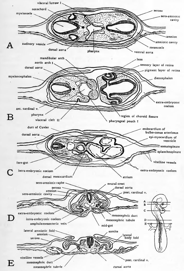 | 705 × 1,034 (188 KB) | S8600021 (talk | contribs) | {{Template:Patten_1920_Figures}} |
You cannot overwrite this file.
File usage
The following 2 pages use this file: