File:Patent ductus arteriosus echocardiogram.jpg

Original file (1,200 × 960 pixels, file size: 133 KB, MIME type: image/jpeg)
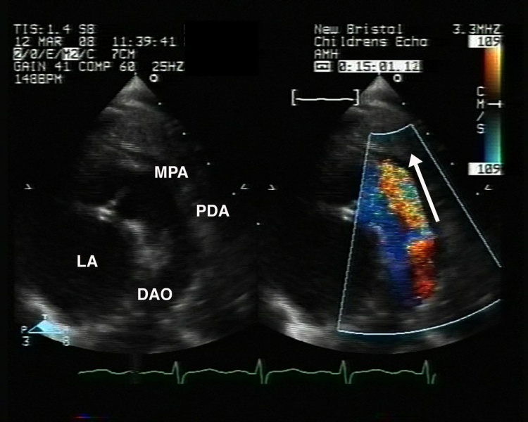
Patent Ductus Arteriosus (PDA) Echocardiogram
Echocardiography, a high parasternal long axis view demonstrating a large patent duct (PDA). Colour flow demonstrates shunting from 'left to right', from the descending aorta (DAO) to the pulmonary arteries (MPA).
- Links: Cardiovascular System - Abnormalities | Patent Ductus Arteriosus cartoon | Patent ductus arteriosus classification | PDA echocardiogram | PDA angiogram
Original file name: 1750-1172-4-17-1.jpg http://www.ojrd.com/content/4/1/17/figure/F1
Reference
<pubmed>19591690</pubmed>| PMC2716300 | Orphanet J Rare Dis.
© 2009 Forsey et al; licensee BioMed Central Ltd.
This is an Open Access article distributed under the terms of the Creative Commons Attribution License (http://creativecommons.org/licenses/by/2.0), which permits unrestricted use, distribution, and reproduction in any medium, provided the original work is properly cited.
File history
Click on a date/time to view the file as it appeared at that time.
| Date/Time | Thumbnail | Dimensions | User | Comment | |
|---|---|---|---|---|---|
| current | 12:40, 27 April 2011 | | 1,200 × 960 (133 KB) | S8600021 (talk | contribs) | ==Patent Ductus Arteriosus (PDA) Echocardiogram== Echocardiography, a high parasternal long axis view demonstrating a large patent duct (PDA). Colour flow demonstrates shunting from 'left to right', from the descending aorta (DAO) to the pulmonary arteri |
You cannot overwrite this file.