File:Patent ductus arteriosus echocardiogram.jpg: Difference between revisions
(==Patent Ductus Arteriosus (PDA) Echocardiogram== Echocardiography, a high parasternal long axis view demonstrating a large patent duct (PDA). Colour flow demonstrates shunting from 'left to right', from the descending aorta (DAO) to the pulmonary arteri) |
mNo edit summary |
||
| (2 intermediate revisions by one other user not shown) | |||
| Line 1: | Line 1: | ||
==Patent Ductus Arteriosus (PDA) Echocardiogram== | ==Patent Ductus Arteriosus (PDA) Echocardiogram== | ||
Echocardiography, a high parasternal long axis view demonstrating a large patent duct (PDA | Echocardiography, a high parasternal long axis view demonstrating a large patent duct (PDA). | ||
Colour flow demonstrates shunting from 'left to right', from the descending aorta (DAO) to the pulmonary arteries (MPA). | |||
:'''Links:''' [[Cardiovascular System - Abnormalities]] | [[:File:Patent Ductus Arteriosus.jpg|Patent Ductus Arteriosus cartoon]] | [[:File:Patent ductus arteriosus classification.jpg|Patent ductus arteriosus classification]] | [[:File:Patent ductus arteriosus echocardiogram.jpg|PDA echocardiogram]] | [[:File:Patent ductus arteriosus angiogram.jpg|PDA angiogram]] | |||
<br> | |||
{{Heart Abnormal}} | |||
<br> | |||
{{Heart Links}} | |||
===Reference=== | |||
<pubmed>19591690</pubmed>| [http://www.ncbi.nlm.nih.gov/pmc/articles/PMC2716300 PMC2716300] | [http://www.ojrd.com/content/4/1/17 Orphanet J Rare Dis.] | |||
====Copyright==== | |||
© 2009 Forsey et al; licensee BioMed Central Ltd. | © 2009 Forsey et al; licensee BioMed Central Ltd. | ||
This is an Open Access article distributed under the terms of the Creative Commons Attribution License (http://creativecommons.org/licenses/by/2.0), which permits unrestricted use, distribution, and reproduction in any medium, provided the original work is properly cited. | This is an Open Access article distributed under the terms of the Creative Commons Attribution License (http://creativecommons.org/licenses/by/2.0), which permits unrestricted use, distribution, and reproduction in any medium, provided the original work is properly cited. | ||
[[Category:Cardiovascular]] [[Category:Ultrasound]] | {{Footer}} | ||
[[Category:Cardiovascular]] [[Category:Ultrasound]] [[Category:Abnormal Development]] | |||
[[Category:Patent Ductus Arteriosus]] | |||
Latest revision as of 10:50, 6 June 2017
Patent Ductus Arteriosus (PDA) Echocardiogram
Echocardiography, a high parasternal long axis view demonstrating a large patent duct (PDA).
Colour flow demonstrates shunting from 'left to right', from the descending aorta (DAO) to the pulmonary arteries (MPA).
- Links: Cardiovascular System - Abnormalities | Patent Ductus Arteriosus cartoon | Patent ductus arteriosus classification | PDA echocardiogram | PDA angiogram
Reference
<pubmed>19591690</pubmed>| PMC2716300 | Orphanet J Rare Dis.
Copyright
© 2009 Forsey et al; licensee BioMed Central Ltd. This is an Open Access article distributed under the terms of the Creative Commons Attribution License (http://creativecommons.org/licenses/by/2.0), which permits unrestricted use, distribution, and reproduction in any medium, provided the original work is properly cited.
Cite this page: Hill, M.A. (2024, April 25) Embryology Patent ductus arteriosus echocardiogram.jpg. Retrieved from https://embryology.med.unsw.edu.au/embryology/index.php/File:Patent_ductus_arteriosus_echocardiogram.jpg
- © Dr Mark Hill 2024, UNSW Embryology ISBN: 978 0 7334 2609 4 - UNSW CRICOS Provider Code No. 00098G
File history
Click on a date/time to view the file as it appeared at that time.
| Date/Time | Thumbnail | Dimensions | User | Comment | |
|---|---|---|---|---|---|
| current | 12:40, 27 April 2011 | 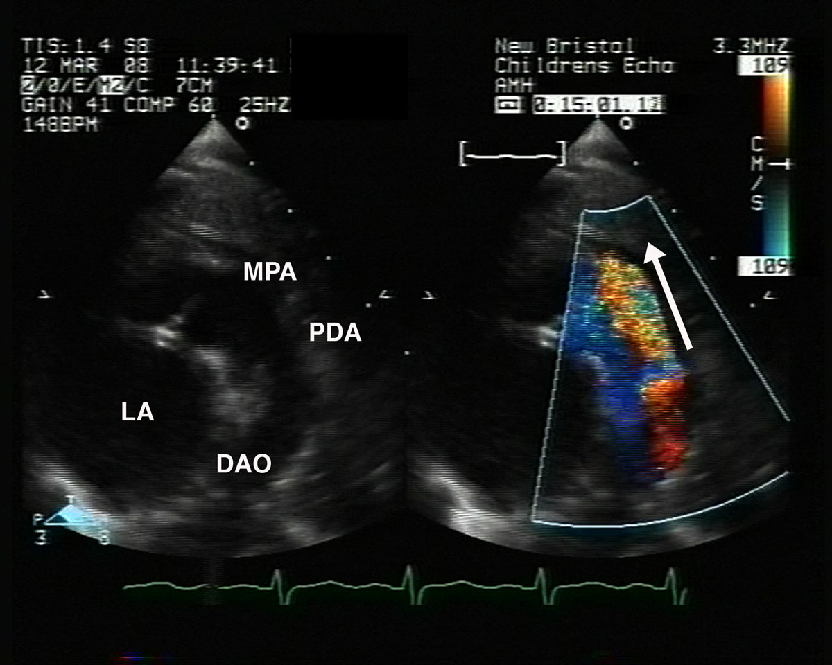 | 1,200 × 960 (133 KB) | S8600021 (talk | contribs) | ==Patent Ductus Arteriosus (PDA) Echocardiogram== Echocardiography, a high parasternal long axis view demonstrating a large patent duct (PDA). Colour flow demonstrates shunting from 'left to right', from the descending aorta (DAO) to the pulmonary arteri |
You cannot overwrite this file.