File:Patent ductus arteriosus classification.jpg: Difference between revisions
From Embryology
mNo edit summary |
|||
| (7 intermediate revisions by 2 users not shown) | |||
| Line 3: | Line 3: | ||
Patent Ductus Arteriosus (PDA) classification system on the basis of angiogram appearance by Krichenko.<ref><pubmed>2929450</pubmed></ref> | Patent Ductus Arteriosus (PDA) classification system on the basis of angiogram appearance by Krichenko.<ref><pubmed>2929450</pubmed></ref> | ||
* '''Type A''' - conical duct with well defined aortic ampulla and constriction near the pulmonary artery end. | |||
* '''Type B''' - large duct with window like structure which is very short in length. | |||
* '''Type C''' - tubular duct without any constriction. | |||
* '''Type D''' - complex duct with multiple constrictions. | |||
* '''Type E''' - elongated duct with constriction remote from the edge of the trachea (as viewed on lateral angiography). | |||
:'''Links:''' [[Cardiovascular_System_-_Patent_Ductus_Arteriosus|Patent Ductus Arteriosus]] | [[Cardiovascular System - Abnormalities]] | [[:File:Patent Ductus Arteriosus.jpg|Patent Ductus Arteriosus cartoon]] | [[:File:Patent ductus arteriosus classification.jpg|Patent ductus arteriosus classification]] | [[:File:Patent ductus arteriosus echocardiogram.jpg|PDA echocardiogram]] | [[:File:Patent ductus arteriosus angiogram.jpg|PDA angiogram]] | |||
The cartoon is based upon several sources.<ref>'''Report of the Review and Implementation Committee for The Report of the Manitoba Pediatric Cardiac Surgery Inquest''' May 2001 image showing heart abnormalities courtesy Government of Manitoba.</ref><ref><pubmed>19591690</pubmed>| [http://www.ncbi.nlm.nih.gov/pmc/articles/PMC2716300 PMC2716300] | [http://www.ojrd.com/content/4/1/17 Orphanet J Rare Dis.]</ref> | |||
<br> | |||
{{Heart Abnormal}} | |||
<br> | |||
{{Heart Links}} | |||
<br> | |||
{{Manitoba Health Report 2001}} | |||
==References== | ==References== | ||
<references/> | <references/> | ||
[[Category:Cardiovascular]] [[Category:Heart]] [[Category:Abnormal Development]] [[Category:Cartoon]] | |||
[[Category:Patent Ductus Arteriosus]] | |||
Latest revision as of 10:49, 6 June 2017
Patent Ductus Arteriosus (PDA) Classification
Patent Ductus Arteriosus (PDA) classification system on the basis of angiogram appearance by Krichenko.[1]
- Type A - conical duct with well defined aortic ampulla and constriction near the pulmonary artery end.
- Type B - large duct with window like structure which is very short in length.
- Type C - tubular duct without any constriction.
- Type D - complex duct with multiple constrictions.
- Type E - elongated duct with constriction remote from the edge of the trachea (as viewed on lateral angiography).
- Links: Patent Ductus Arteriosus | Cardiovascular System - Abnormalities | Patent Ductus Arteriosus cartoon | Patent ductus arteriosus classification | PDA echocardiogram | PDA angiogram
The cartoon is based upon several sources.[2][3]
| Heart Abnormal: Tutorial Abnormalities | atrial septal defects | double outlet right ventricle | hypoplastic left heart | patent ductus arteriosus | transposition of the great vessels | Tetralogy of Fallot | ventricular septal defects | coarctation of the aorta | Category ASD | Category PDA | Category ToF | Category VSD | ICD10 - Cardiovascular | ICD11 |
Image source: Report of the Review and Implementation Committee for The Report of the Manitoba Pediatric Cardiac Surgery Inquest May 2001 image showing heart abnormalities courtesy Government of Manitoba.
References
- ↑ <pubmed>2929450</pubmed>
- ↑ Report of the Review and Implementation Committee for The Report of the Manitoba Pediatric Cardiac Surgery Inquest May 2001 image showing heart abnormalities courtesy Government of Manitoba.
- ↑ <pubmed>19591690</pubmed>| PMC2716300 | Orphanet J Rare Dis.
File history
Click on a date/time to view the file as it appeared at that time.
| Date/Time | Thumbnail | Dimensions | User | Comment | |
|---|---|---|---|---|---|
| current | 13:46, 27 April 2011 | 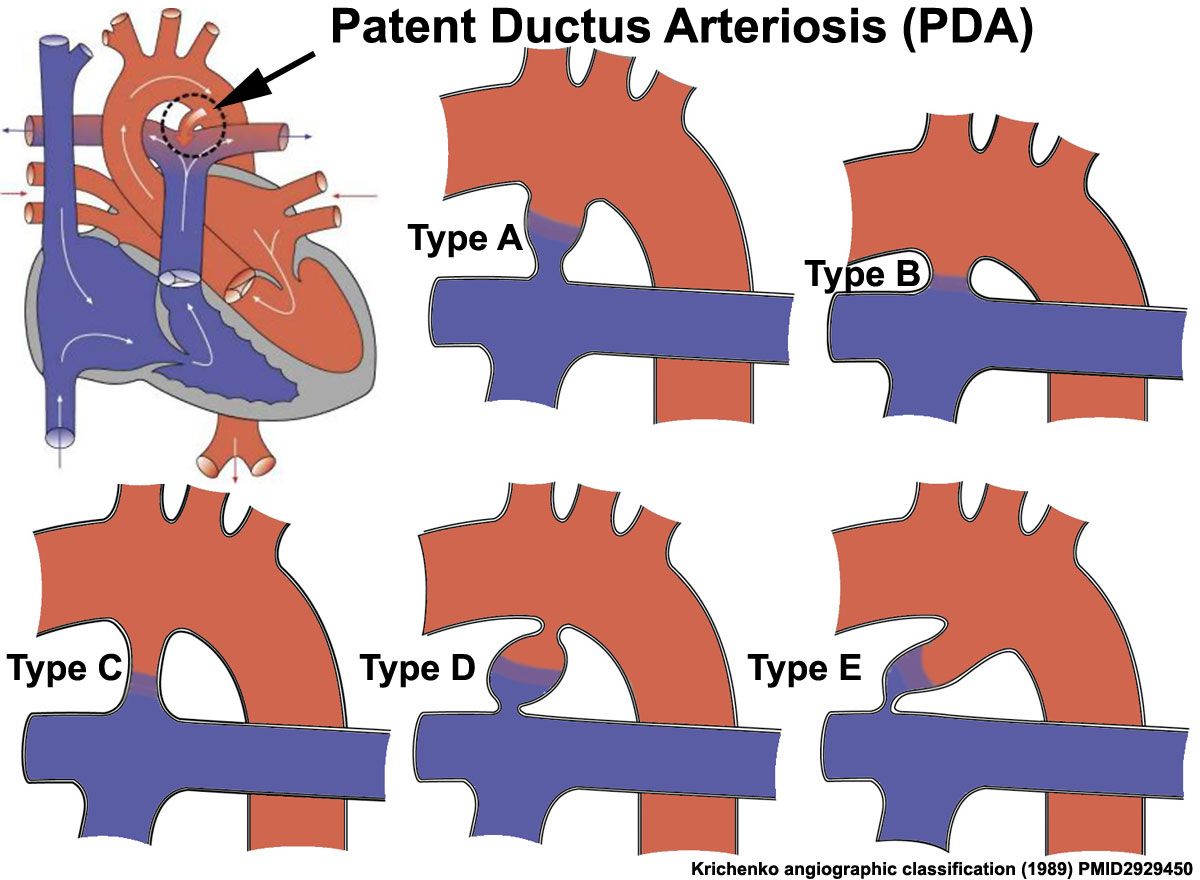 | 1,200 × 880 (138 KB) | S8600021 (talk | contribs) | ==Patent Ductus Arteriosus (PDA) Classification== Patent Ductus Arteriosus (PDA) classification system on the basis of angiogram appearance by Krichenko.<ref><pubmed>2929450</pubmed></ref> {{Template:Manitoba Health Report 2001}} <pubmed>19591690</pu |
You cannot overwrite this file.