File:Paramesonephric ducts.jpg: Difference between revisions
From Embryology
No edit summary |
mNo edit summary |
||
| (3 intermediate revisions by the same user not shown) | |||
| Line 1: | Line 1: | ||
==Paramesonephric Ducts== | |||
[[:File:Gray1109.jpg|Gray's Anatomy - Fig. 1109]] | |||
Urogenital sinus of female human embryo of eight and a half to nine weeks old (from model by Keibel) | Urogenital sinus of female human embryo of eight and a half to nine weeks old (from model by Keibel) | ||
(Image: Gray's Anatomy) | (Image: Gray's Anatomy) | ||
:'''Links:''' [[Uterus Development]] | [[:File:Gray1109.jpg|Gray's Figure 1109]] | |||
{{Genital Links}} | |||
{{Footer}} | |||
[[Category:Uterus]] [[Category:Genital]] [[Category:Gray's 1918 Anatomy]] [[Category:Cartoon]] | [[Category:Uterus]] [[Category:Genital]] [[Category:Gray's 1918 Anatomy]] [[Category:Cartoon]] | ||
Latest revision as of 14:06, 17 March 2016
Paramesonephric Ducts
Urogenital sinus of female human embryo of eight and a half to nine weeks old (from model by Keibel)
(Image: Gray's Anatomy)
- Links: Uterus Development | Gray's Figure 1109
Cite this page: Hill, M.A. (2024, April 19) Embryology Paramesonephric ducts.jpg. Retrieved from https://embryology.med.unsw.edu.au/embryology/index.php/File:Paramesonephric_ducts.jpg
- © Dr Mark Hill 2024, UNSW Embryology ISBN: 978 0 7334 2609 4 - UNSW CRICOS Provider Code No. 00098G
File history
Click on a date/time to view the file as it appeared at that time.
| Date/Time | Thumbnail | Dimensions | User | Comment | |
|---|---|---|---|---|---|
| current | 22:32, 12 April 2010 | 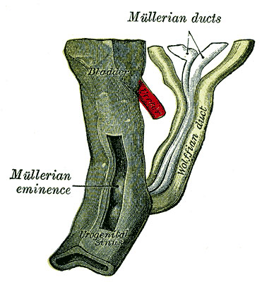 | 371 × 400 (43 KB) | S8600021 (talk | contribs) | Urogenital sinus of female human embryo of eight and a half to nine weeks old (from model by Keibel) (Image: Gray's Anatomy) Category:Uterus Category:Genital Category:Gray's 1918 Anatomy Category:Cartoon |
You cannot overwrite this file.
File usage
The following 4 pages use this file: