File:Pancreatic islet.png
Pancreatic_islet.png (600 × 462 pixels, file size: 425 KB, MIME type: image/png)
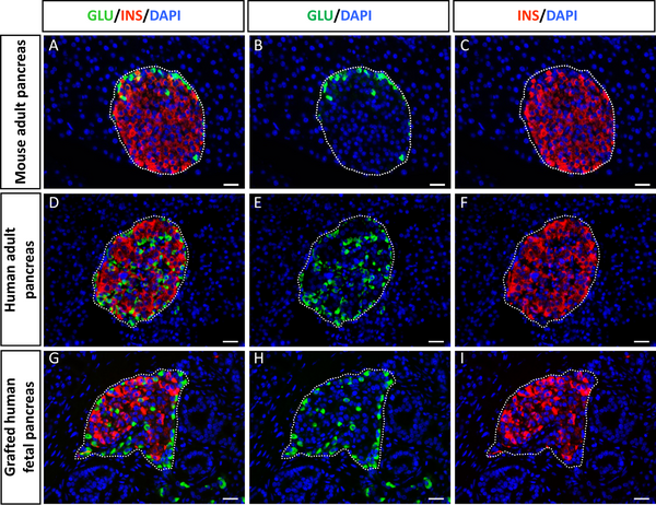
Figure 1. Cytoarchitecture of newly formed islets from human fetal pancreas.
A–C: Section of adult mouse pancreas stained for glucagon (green) and insulin (red).
D–F: Section of an adult human pancreas stained for glucagon (green) and insulin (red).
G–I: Section of a human fetal pancreas analyzed 4.5 months after transplantation and stained for glucagon (green) and insulin (red). Nuclear staining (blue) was performed with DAPI.
Scale bars: 25 µm.
Journal.pone.0003559.g001.png
Beta cells within single human islets originate from multiple progenitors. Scharfmann R, Xiao X, Heimberg H, Mallet J, Ravassard P. PLoS One. 2008;3(10):e3559. Epub 2008 Oct 29. PMID: 18958289
Citation: Scharfmann R, Xiao X, Heimberg H, Mallet J, Ravassard P (2008) Beta Cells within Single Human Islets Originate from Multiple Progenitors. PLoS ONE 3(10): e3559. doi:10.1371/journal.pone.0003559
Editor: Tailoi Chan-Ling, University of Sydney, Australia
Received: February 5, 2008; Accepted: October 9, 2008; Published: October 29, 2008
Copyright: © 2008 Scharfmann et al. This is an open-access article distributed under the terms of the Creative Commons Attribution License, which permits unrestricted use, distribution, and reproduction in any medium, provided the original author and source are credited.
File history
Click on a date/time to view the file as it appeared at that time.
| Date/Time | Thumbnail | Dimensions | User | Comment | |
|---|---|---|---|---|---|
| current | 19:49, 5 October 2009 | | 600 × 462 (425 KB) | S8600021 (talk | contribs) | Figure 1. Cytoarchitecture of newly formed islets from human fetal pancreas. A–C: Section of adult mouse pancreas stained for glucagon (green) and insulin (red). D–F: Section of an adult human pancreas stained for glucagon (green) and insulin (red) |
You cannot overwrite this file.