File:Pancreatic islet.png: Difference between revisions
No edit summary |
mNo edit summary |
||
| (4 intermediate revisions by 2 users not shown) | |||
| Line 1: | Line 1: | ||
==Pancreatic Islet - Endocrine Cells== | |||
Cytoarchitecture of newly formed islets from human fetal pancreas | |||
A–C: Section of adult mouse pancreas stained for glucagon (green) and insulin (red). | '''A–C:''' Section of adult mouse pancreas stained for glucagon (green) and insulin (red). | ||
D–F: Section of an adult human pancreas stained for glucagon (green) and insulin (red). | '''D–F:''' Section of an adult human pancreas stained for glucagon (green) and insulin (red). | ||
G–I: Section of a human fetal pancreas analyzed 4.5 months after transplantation and stained for glucagon (green) and insulin (red). Nuclear staining (blue) was performed with DAPI. | '''G–I:''' Section of a human fetal pancreas analyzed 4.5 months after transplantation and stained for glucagon (green) and insulin (red). Nuclear staining (blue) was performed with DAPI. | ||
Scale bars: 25 µm. | Scale bars: 25 µm. | ||
'''Links:''' [[:File:Human- pancreatic adult islet.jpg|Islet]] | [[:File:Human- pancreatic adult islet-insulin.jpg|Insulin]] | [[:File:Human-_pancreatic_adult_islet-glucagon.jpg|Glucagon]] | [[:File:Pancreatic_islet.png|original image panel]] | {{pancreas}} | |||
===Reference=== | |||
{{#pmid:18958289}} | |||
Copyright | ====Copyright==== | ||
© 2008 Scharfmann et al. This is an open-access article distributed under the terms of the Creative Commons Attribution License, which permits unrestricted use, distribution, and reproduction in any medium, provided the original author and source are credited. | |||
Original File name: Figure 1. Journal.pone.0003559.g001.png | |||
{{Footer}} | |||
[[Category:Endocrine]] [[Category:Pancreas]] | [[Category:Endocrine]] [[Category:Pancreas]] | ||
Latest revision as of 10:26, 31 July 2019
Pancreatic Islet - Endocrine Cells
Cytoarchitecture of newly formed islets from human fetal pancreas
A–C: Section of adult mouse pancreas stained for glucagon (green) and insulin (red).
D–F: Section of an adult human pancreas stained for glucagon (green) and insulin (red).
G–I: Section of a human fetal pancreas analyzed 4.5 months after transplantation and stained for glucagon (green) and insulin (red). Nuclear staining (blue) was performed with DAPI.
Scale bars: 25 µm.
Links: Islet | Insulin | Glucagon | original image panel | pancreas
Reference
Scharfmann R, Xiao X, Heimberg H, Mallet J & Ravassard P. (2008). Beta cells within single human islets originate from multiple progenitors. PLoS ONE , 3, e3559. PMID: 18958289 DOI.
Copyright
© 2008 Scharfmann et al. This is an open-access article distributed under the terms of the Creative Commons Attribution License, which permits unrestricted use, distribution, and reproduction in any medium, provided the original author and source are credited.
Original File name: Figure 1. Journal.pone.0003559.g001.png
Cite this page: Hill, M.A. (2024, April 19) Embryology Pancreatic islet.png. Retrieved from https://embryology.med.unsw.edu.au/embryology/index.php/File:Pancreatic_islet.png
- © Dr Mark Hill 2024, UNSW Embryology ISBN: 978 0 7334 2609 4 - UNSW CRICOS Provider Code No. 00098G
File history
Click on a date/time to view the file as it appeared at that time.
| Date/Time | Thumbnail | Dimensions | User | Comment | |
|---|---|---|---|---|---|
| current | 19:49, 5 October 2009 | 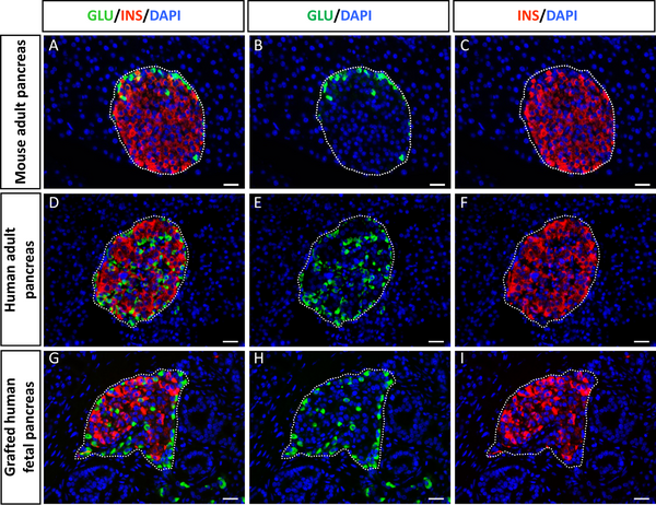 | 600 × 462 (425 KB) | S8600021 (talk | contribs) | Figure 1. Cytoarchitecture of newly formed islets from human fetal pancreas. A–C: Section of adult mouse pancreas stained for glucagon (green) and insulin (red). D–F: Section of an adult human pancreas stained for glucagon (green) and insulin (red) |
You cannot overwrite this file.