File:Pancreatic duct developing.jpg
Pancreatic_duct_developing.jpg (400 × 322 pixels, file size: 15 KB, MIME type: image/jpeg)
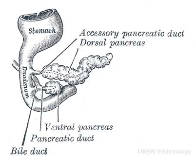
Fig. 1101. Pancreas of a human embryo of five weeks
(Kollmann)
Pancreatic Duct
The initial formation of the pancreas as two separate lobes each with their own duct that fuses leads a range of anatomical variations in the adult exocrine pancreatic duct. Pancreatic duct five variation classification: common, ansa pancreatica, branch fusion, looped, and separated. Accessory pancreatic duct (APD, of Santorini) in the embryo is the main drainage duct of the dorsal pancreatic bud emptying into the minor duodenal papilla. In the adult it has been further classified as either long-type (joins main pancreatic duct at pancreas neck portion) and short-type (joins main pancreatic duct near first inferior branch).
Main Pancreatic Duct
- (MPD or Wirsung's duct) forms within the dorsal pancreatic bud and is present in the body and tail of the pancreas.
- Discovered by Johann Georg Wirsung (1589 - 1643) a German physician who worked as a prosector in Padua.
Accessory Pancreatic Duct
- (APD or Santorini’s duct) is present mainly in the head of the pancreas.
- Originally dissected and delineated by Giovanni Domenico Santorini (1681 - 1737) an Italian anatomist.
- Endoscopic Retrograde Cholangiopancreatography (ERCP) is a medical procedure which allows an injected dye to display the duct system on an x ray (pancreatograms).
File history
Click on a date/time to view the file as it appeared at that time.
| Date/Time | Thumbnail | Dimensions | User | Comment | |
|---|---|---|---|---|---|
| current | 11:18, 4 October 2009 | | 400 × 322 (15 KB) | S8600021 (talk | contribs) |
You cannot overwrite this file.
File usage
The following 15 pages use this file:
- 2009 Lecture 20
- 2010 Lecture 19
- 2011 Lab 5 - Late Embryo
- ANAT2341 Lab 5 - Fetal
- ANAT2341 Lab 5 - Late Embryo
- BGDB Gastrointestinal - Activity 3
- BGDB Gastrointestinal - Late Embryo
- BGD Lecture - Endocrine Development
- BGD Lecture - Gastrointestinal System Development
- Endocrine - Pancreas Development
- Endocrine System Development
- Gastrointestinal Tract - Pancreas Development
- Lecture - Endocrine Development
- Lecture - Gastrointestinal Development 2013
- P