File:Ovary corpus luteum.jpg
From Embryology

Size of this preview: 800 × 418 pixels. Other resolution: 2,178 × 1,137 pixels.
Original file (2,178 × 1,137 pixels, file size: 376 KB, MIME type: image/jpeg)
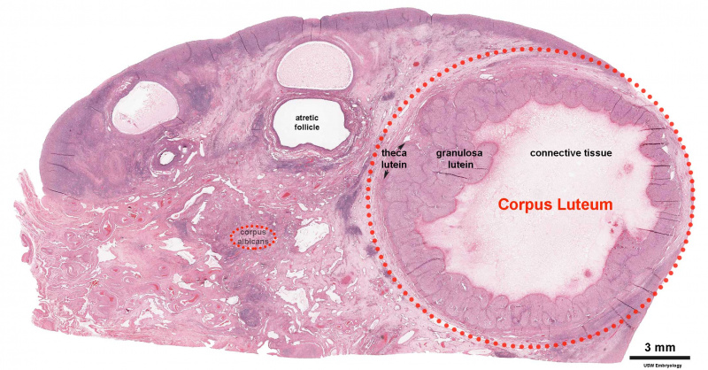
Ovary showing corpus luteum and atretic follicles.
Histological image, H&E stained.
Image Source: UNSW Embryology http://embryology.med.unsw.edu.au/Medicine/BGDlabfertilization6.htm
File history
Click on a date/time to view the file as it appeared at that time.
| Date/Time | Thumbnail | Dimensions | User | Comment | |
|---|---|---|---|---|---|
| current | 19:19, 5 May 2018 | | 2,178 × 1,137 (376 KB) | Z8600021 (talk | contribs) | |
| 18:58, 5 May 2018 |  | 2,178 × 1,137 (375 KB) | Z8600021 (talk | contribs) | ||
| 18:50, 5 May 2018 | 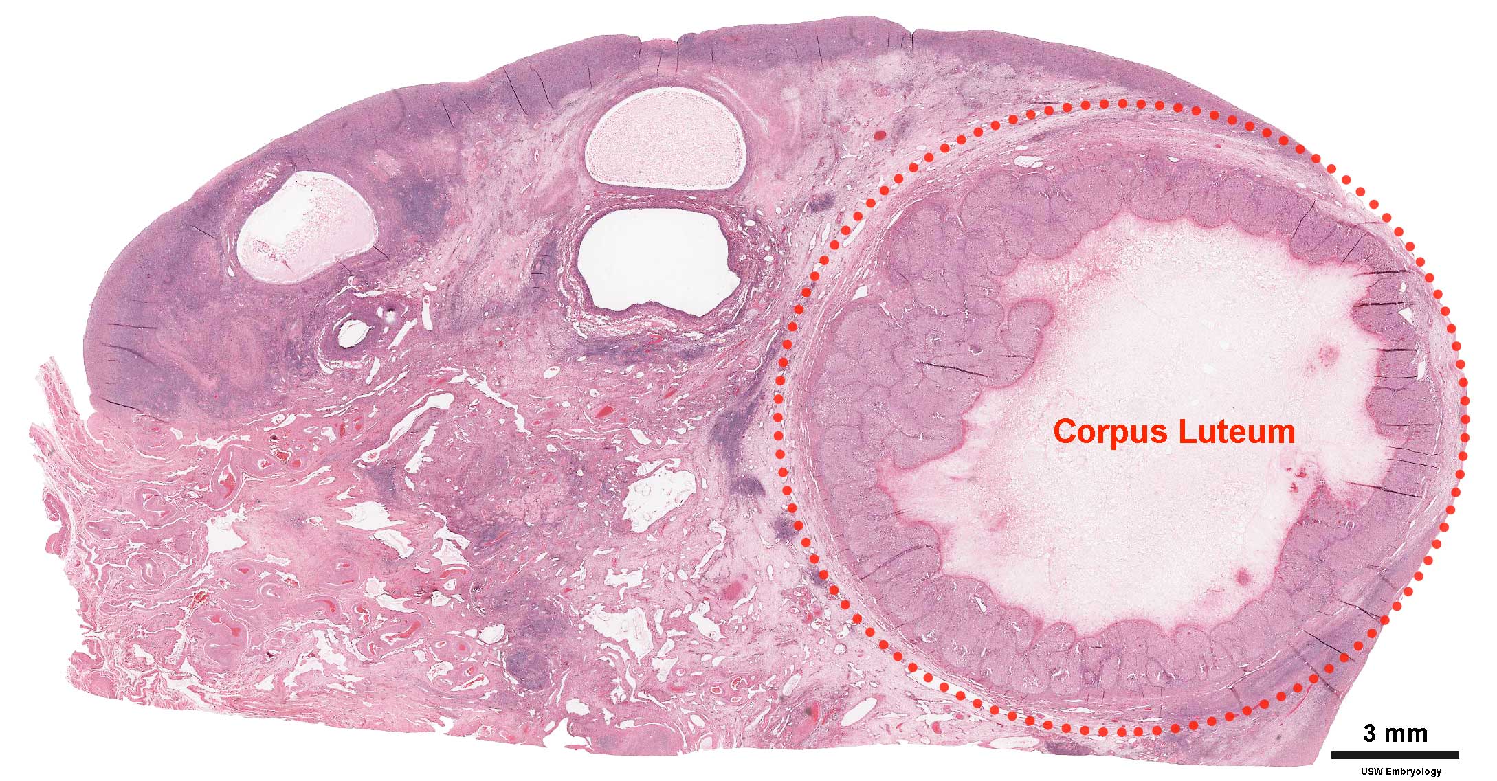 | 2,178 × 1,137 (369 KB) | Z8600021 (talk | contribs) | ||
| 10:10, 3 August 2009 |  | 800 × 455 (103 KB) | MarkHill (talk | contribs) | Ovary showing corpus luteum and atretic follicles. Histological image, H&E stained. Image Source: UNSW Embryology http://embryology.med.unsw.edu.au/Medicine/BGDlabfertilization6.htm |
You cannot overwrite this file.
File usage
The following 17 pages use this file:
- 2009 Lecture 3
- 2010 BGD Practical 3 - Implantation
- 2010 Foundations Lecture - Introduction to Human Development
- 2010 Lecture 3
- 2011 Lab 2 - Week 2
- ANAT2241 Female Reproductive System
- ANAT2341 Lab 2 - Week 2
- BGDA Practical - Female Reproductive Tract Histology
- BGDA Practical 3 - Implantation
- Corpus Luteum Development
- Foundations Lecture - Introduction to Human Development
- Foundations Practical - Week 1 and 2
- Lecture - Week 1 and 2 Development
- Menstrual Cycle
- Ovary Development
- Pre-Medicine Program - Embryology
- Talk:2011 Lab 2 - Week 2