|
|
| Line 20: |
Line 20: |
| |} | | |} |
| ===Reference=== | | ===Reference=== |
| <pubmed>23409002</pubmed>| [http://www.plosone.org/article/info%3Adoi%2F10.1371%2Fjournal.pone.0055578 PLoS One.]
| | {{#pmid:23409002}} |
|
| |
|
| ====Copyright==== | | ====Copyright==== |
| Line 28: |
Line 28: |
| Fig. 7 doi:10.1371/journal.pone.0055578.g007 Modified in size and labelling. | | Fig. 7 doi:10.1371/journal.pone.0055578.g007 Modified in size and labelling. |
|
| |
|
| | | {{Footer}} |
| [[Category:Ovary]][[Category:Bovine]][[Category:Cartoon]] | | [[Category:Ovary]][[Category:Bovine]][[Category:Cartoon]] |
Latest revision as of 10:03, 27 March 2020
Ovarian Development Model
- A - The development of the ovary commences at the mesonephric surface epithelium (yellow cells) in the location of the future gonadal ridge.
- B - Some mesonephric surface epithelial cells change phenotype into GREL (Gonadal Ridge Epithelial-Like) cells (yellow-blue cells).
- C - The GREL cells proliferate and the basal lamina underlying the mesonephric surface epithelium breaks down allowing stromal cells (green) to penetrate into the gonadal ridge.
- D - GREL cells continue to proliferate and PGCs (grey) migrate into the ridge between the GREL cells. Mesonephric stroma including vasculature (red) continues to penetrate and expand in the ovary.
- E - Oogonia proliferate and stroma penetrates further towards the ovarian surface enclosing oogonia and GREL cells into ovigerous cords. The cords are surrounded by a basal lamina at their interface with stroma, but are open to the ovarian surface. Stromal areas including those between the ovigerous cords contain capillaries.
- F - A compartmentalization into cortex and medulla becomes obvious. The cortex is characterised by alternating areas of ovigerous cords and stroma, whereas the medulla is formed by stromal cells, vasculature and tubules originating from the mesonephros (rete ovarii). Once stroma penetrates below the cells on the surface it spreads laterally. The GREL cells at the surface are then aligned by a basal lamina at their interface with the stroma and begin to differentiate into typical ovarian surface epithelium (yellow cells). Some germ cells at the surface are also compartmentalized to the surface as stroma expands below it.
- G - Ovigerous cords are partitioned into smaller cords and eventually into follicles. These contain GREL cells that form granulosa cells (blue cells) and oogonia that form oocytes. The first primordial follicles appear in the inner cortex-medulla region, surrounded by a basal lamina. A now fully intact basal lamina underlies multiple layers of surface epithelial cells.
- H - At the final stage the surface epithelium becomes mostly single-layered and a tunica albuginea, densely packed with fibres, develops from the stroma below the surface epithelial basal lamina. Some primordial follicles become activated and commence development into primary and preantral follicles.
- Links: Ovary Development | Bovine Development
|
- GREL - Gonadal Ridge Epithelial-Like (yellow-blue cells)
- PGC - Primordial Germ Cell (grey)
- Yellow cells - mesonephric surface epithelium
|
Reference
Hummitzsch K, Irving-Rodgers HF, Hatzirodos N, Bonner W, Sabatier L, Reinhardt DP, Sado Y, Ninomiya Y, Wilhelm D & Rodgers RJ. (2013). A new model of development of the mammalian ovary and follicles. PLoS ONE , 8, e55578. PMID: 23409002 DOI.
Copyright
© 2013 Hummitzsch et al. This is an open-access article distributed under the terms of the Creative Commons Attribution License, which permits unrestricted use, distribution, and reproduction in any medium, provided the original author and source are credited.
Fig. 7 doi:10.1371/journal.pone.0055578.g007 Modified in size and labelling.
Cite this page: Hill, M.A. (2024, April 19) Embryology Ovarian development model.jpg. Retrieved from https://embryology.med.unsw.edu.au/embryology/index.php/File:Ovarian_development_model.jpg
- What Links Here?
- © Dr Mark Hill 2024, UNSW Embryology ISBN: 978 0 7334 2609 4 - UNSW CRICOS Provider Code No. 00098G
Click on a date/time to view the file as it appeared at that time.
| Date/Time | Thumbnail | Dimensions | User | Comment |
|---|
| current | 12:35, 5 November 2014 | 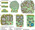 | 1,200 × 1,036 (445 KB) | Z8600021 (talk | contribs) | ===Reference=== <pubmed>23409002</pubmed>| [http://www.plosone.org/article/info%3Adoi%2F10.1371%2Fjournal.pone.0055578 PLoS One.] ====Copyright==== © 2013 Hummitzsch et al. This is an open-access article distributed under the terms of the Creative C... |
You cannot overwrite this file.
The following 2 pages use this file:
This file contains additional information, probably added from the digital camera or scanner used to create or digitise it.
If the file has been modified from its original state, some details may not fully reflect the modified file.
