File:Ossification endochondral 1.jpg: Difference between revisions
From Embryology
No edit summary |
|||
| (7 intermediate revisions by 2 users not shown) | |||
| Line 1: | Line 1: | ||
Endochondral Ossification | ==Endochondral Ossification== | ||
Histological image of a developing vertebra and intervertebral disc ({{rat}}), scale bar 80 microns. | |||
* vertebra - cartilage template and developing bony collar (top of image) | |||
* intervertebral disc - nucleus pulposus and annular fibrocartilage (bottom of image) | |||
See also adjacent region image of [[:File:Ossification_endochondral_01.jpg|Developing Vertebra]] | |||
===Development=== | |||
* Intervertebral disc nucleus pulposus - derived from the {{notochord}} | |||
* Vertebra - derived from the sclerotome of the paired somites. | |||
:'''Links:''' {{axial skeleton}} | [[:File:Ossification endochondral 1.jpg|Image - Intervertebral Disc]] | [[:File:Ossification_endochondral_01.jpg|Image - Vertebra]] | {{bone}} | [[Cartilage Histology]] | [[Bone Histology]] | |||
{{Footer}} | |||
== Image version links == | == Image version links == | ||
| Line 6: | Line 26: | ||
[[:File:Ossification endochondral 1b.jpg|Medium 600px]] | [[:File:Ossification endochondral 1c.jpg|Small 400px]] | [[:File:Ossification endochondral 1b.jpg|Medium 600px]] | [[:File:Ossification endochondral 1c.jpg|Small 400px]] | ||
Original File Name: Endochondral9x10n3-1000px.jpg | |||
Image Source: UNSW Embryology | |||
[[Category:Histology]] [[Category:Musculoskeletal]] | [[Category:Histology]] [[Category:Musculoskeletal]] [[Category:Notochord]] [[Category:Cartilage]] [[Category:Bone]] [[Category:Axial Skeleton]] | ||
Latest revision as of 19:34, 2 May 2018
Endochondral Ossification
Histological image of a developing vertebra and intervertebral disc (rat), scale bar 80 microns.
- vertebra - cartilage template and developing bony collar (top of image)
- intervertebral disc - nucleus pulposus and annular fibrocartilage (bottom of image)
See also adjacent region image of Developing Vertebra
Development
- Intervertebral disc nucleus pulposus - derived from the notochord
- Vertebra - derived from the sclerotome of the paired somites.
- Links: axial skeleton | Image - Intervertebral Disc | Image - Vertebra | bone | Cartilage Histology | Bone Histology
Cite this page: Hill, M.A. (2024, April 16) Embryology Ossification endochondral 1.jpg. Retrieved from https://embryology.med.unsw.edu.au/embryology/index.php/File:Ossification_endochondral_1.jpg
- © Dr Mark Hill 2024, UNSW Embryology ISBN: 978 0 7334 2609 4 - UNSW CRICOS Provider Code No. 00098G
Image version links
Large 1000px | 800px | Medium 600px | Small 400px
Original File Name: Endochondral9x10n3-1000px.jpg
Image Source: UNSW Embryology
File history
Click on a date/time to view the file as it appeared at that time.
| Date/Time | Thumbnail | Dimensions | User | Comment | |
|---|---|---|---|---|---|
| current | 10:53, 14 September 2009 | 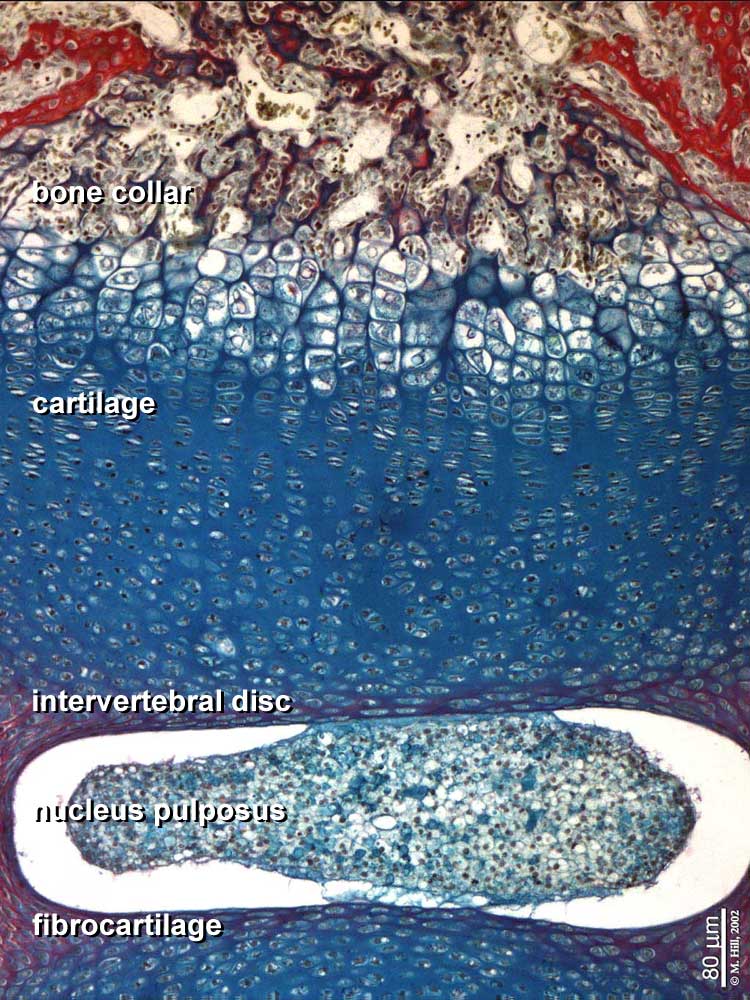 | 750 × 1,000 (147 KB) | S8600021 (talk | contribs) | Endochondral9x10n3-1000px.jpg |
You cannot overwrite this file.
File usage
The following page uses this file: