File:Ninian1939 plate01.jpg

Original file (1,280 × 697 pixels, file size: 171 KB, MIME type: image/jpeg)
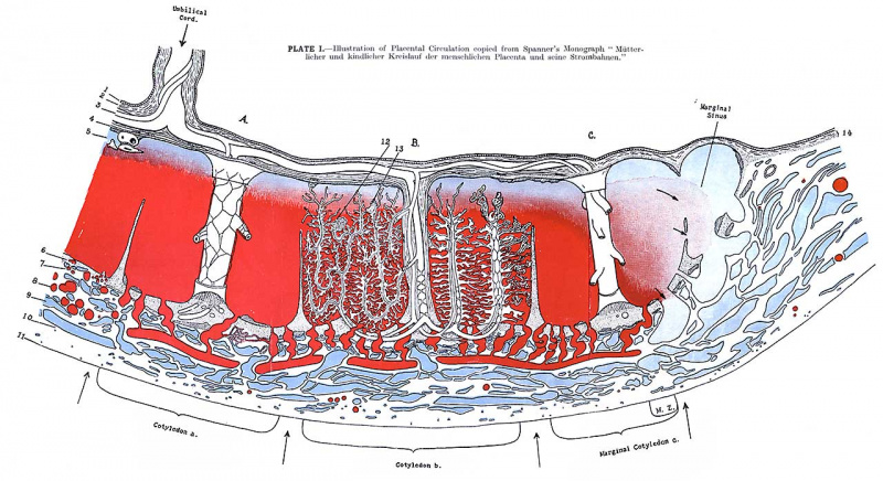
Plate 1. Illustration of Placental Circulation
Copied from Spanner’s Monograph “Mutterlicher und kiderlicher Kreislauf der Menschlichen und seine Strombahnen.” (Some details included in his illustration are omitted).
l. Amnion epithelium. 2-4. Membrana chorii (Chorion plate). 2. Chorion connective tissue 3. Foetal artery and vein. 4. Chorial trophoblast (or its derivatives). Sub-murial blood space. 6. Nitabach’s fibrinoid layer. 7. Decidua basalis with many sections of utero-placental whirl artery. 8. Maternal artery. 9. Maternal vein. 10. Muscularis uteri. ll. Peritoneum. 12. White infarct. (fibrinoid knot). 13. Cell island (trophoblast). l-1. Chorion laeve. A. B. C. Three big anchoring villous trunks to which the Cotyledons a. and b., limited by septs (4 lower arrows), correspond as well as Marginal Cotyledon c. .\I.Z. Marginal Zone.
cotyledon b. — Shows Spanner’s conception of the branching of the villous tree. The outstanding characteristic of the branching of the villous tree is that the smaller villous trunks run in the form of recurrent loops parallel to each other, running vertically from the basal limits of the placenta towards the chorion. This arrangement of the villous tree brings to mind the picture of an antique chandelier and its candles. Note that the main vessels of the villous trunk (B) run at first into the trophoblast of the basal plate, turn there and enter (in the form of three smaller recurrent trunks) the intervillous space. In addition from the centre of villous trunk (B) comes a minor villous trunk which also runs basalwards and is connected by small anchoring villi with the basal plate but finally turns towards the chorion.
Grateful Acknowledgement is made to Professor Spanner for permission to reproduce this coloured plate.
Reference
Ninian F. Circulation of the maternal blood through the placenta. (1939) Irish J. Med. Sci. 14(2): 59-65.
Cite this page: Hill, M.A. (2024, April 24) Embryology Ninian1939 plate01.jpg. Retrieved from https://embryology.med.unsw.edu.au/embryology/index.php/File:Ninian1939_plate01.jpg
- © Dr Mark Hill 2024, UNSW Embryology ISBN: 978 0 7334 2609 4 - UNSW CRICOS Provider Code No. 00098G
File history
Click on a date/time to view the file as it appeared at that time.
| Date/Time | Thumbnail | Dimensions | User | Comment | |
|---|---|---|---|---|---|
| current | 21:39, 6 June 2018 | | 1,280 × 697 (171 KB) | Z8600021 (talk | contribs) | |
| 21:23, 6 June 2018 |  | 3,750 × 2,550 (824 KB) | Z8600021 (talk | contribs) | {{Ref-Ninian1939}} |
You cannot overwrite this file.
File usage
The following page uses this file: