File:Ngn1 and Ngn2 in DRG development.png
From Embryology
No higher resolution available.
Ngn1_and_Ngn2_in_DRG_development.png (384 × 557 pixels, file size: 212 KB, MIME type: image/png)
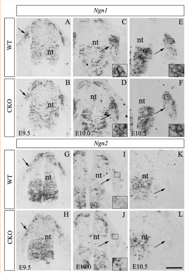
This figure compares of the concentration of cells expressing Ngn-1 and Ngn-2 in the DRG between wild-type and Rbpj knock out mice at E10.0 and E10.5. This figure demonstrates that loss of Rbpj signalling function does not affect neurogenin activity either in the migratory phase of neural crest cells at E9.5 and E10.0 or in post-migratory phase of neural crest cells at E10.0 and E10.5 within the DRG. Arrows in point to a cluster of migrating NCCs or to post-migratory NCCs condensed in the DRG.
File history
Click on a date/time to view the file as it appeared at that time.
| Date/Time | Thumbnail | Dimensions | User | Comment | |
|---|---|---|---|---|---|
| current | 00:47, 15 October 2018 | | 384 × 557 (212 KB) | Z5229597 (talk | contribs) | This figure compares of the concentration of cells expressing Ngn-1 and Ngn-2 in the DRG between wild-type and Rbpj knock out mice at E10.0 and E10.5. This figure demonstrates that loss of Rbpj function does not affect neurogenin activity either in the... |
You cannot overwrite this file.
File usage
The following 2 pages use this file: