File:Neuron cartoon.jpg
From Embryology
No higher resolution available.
Neuron_cartoon.jpg (469 × 500 pixels, file size: 11 KB, MIME type: image/jpeg)
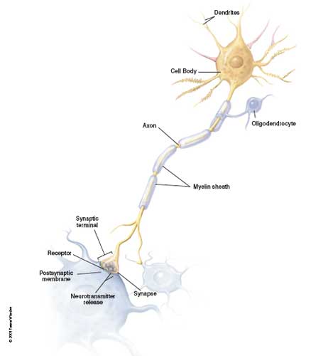
Neuron cartoon
This simplified diagram of a single neuron and its associated glial cell from the central nervous system identifies key cellular structures.
The human brain contains about 86 billion neurons and about the same number of non-neuronal cells. PMID 22723358
Reference
NIH USA Original File name: Neuron1.jpg
Cite this page: Hill, M.A. (2024, April 25) Embryology Neuron cartoon.jpg. Retrieved from https://embryology.med.unsw.edu.au/embryology/index.php/File:Neuron_cartoon.jpg
- © Dr Mark Hill 2024, UNSW Embryology ISBN: 978 0 7334 2609 4 - UNSW CRICOS Provider Code No. 00098G
File history
Click on a date/time to view the file as it appeared at that time.
| Date/Time | Thumbnail | Dimensions | User | Comment | |
|---|---|---|---|---|---|
| current | 15:01, 10 August 2009 | | 469 × 500 (11 KB) | MarkHill (talk | contribs) | Neuron cartoon Original File name: Neuron1.jpg Source: NIH USA |
You cannot overwrite this file.