File:Neuroacanthocytosis.jpg
Neuroacanthocytosis.jpg (487 × 584 pixels, file size: 91 KB, MIME type: image/jpeg)
Neuroacanthocytosis
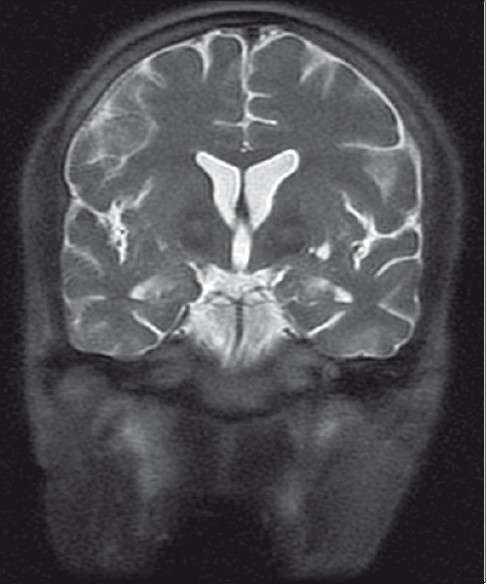
Figure 2
Coronal T2-weighted images showing features as described in Figure 1
(Figure 1
T2-weighted image is showing symmetrical hyperintense signal changes in anterior medial globus pallidus with surrounding hypointensity in the globus pallidus. These imaging features have been termed the “eye-of-the-tiger” sign)
Reference
<pubmed>21716872</pubmed>
This is an open-access article distributed under the terms of the Creative Commons Attribution-Noncommercial-Share Alike 3.0 Unported, which permits unrestricted use, distribution, and reproduction in any medium, provided the original work is properly cited.
Assessment
+ Image is relevant to group topic. - Figure includes no explanation just the original legend. Lacks peer teaching component. For example, What is Neuroacanthocytosis and what is the method of imaging being used? - I had earlier asked for a better legend, it does not appear to be any better. Mark Hill 14:57, 20 September 2011 (EST) Is this figure 2 or 1? Your legend description makes no sense. Please fix this, I have already fixed your referencing. + Copyright, citation and student disclaimer included.
- Note - This image was originally uploaded as part of a student project and may contain inaccuracies in either description or acknowledgements. Students have been advised in writing concerning the reuse of content and may accidentally have misunderstood the original terms of use. If image reuse on this non-commercial educational site infringes your existing copyright, please contact the site editor for immediate removal.
Cite this page: Hill, M.A. (2024, April 19) Embryology Neuroacanthocytosis.jpg. Retrieved from https://embryology.med.unsw.edu.au/embryology/index.php/File:Neuroacanthocytosis.jpg
- © Dr Mark Hill 2024, UNSW Embryology ISBN: 978 0 7334 2609 4 - UNSW CRICOS Provider Code No. 00098G
File history
Click on a date/time to view the file as it appeared at that time.
| Date/Time | Thumbnail | Dimensions | User | Comment | |
|---|---|---|---|---|---|
| current | 14:25, 20 September 2011 | | 487 × 584 (91 KB) | Z3290379 (talk | contribs) | '''Figure 2''' Coronal T2-weighted images showing features as described in Figure 1 (Figure 1 T2-weighted image is showing symmetrical hyperintense signal changes in anterior medial globus pallidus with surrounding hypointensity in the globus pallidus |
You cannot overwrite this file.
File usage
The following 2 pages use this file: