File:Neural Crest Migration.png
From Embryology
No higher resolution available.
Neural_Crest_Migration.png (363 × 144 pixels, file size: 107 KB, MIME type: image/png)
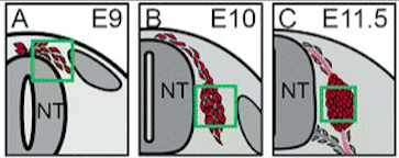
Illustration of a transverse section of the neural tube at E9, E10 and E11.5. The cells that contribute to the DRG are labeled in red.
Reference
Gay MH, Valenta T, Herr P, Paratore-Hari L, Basler K & Sommer L. (2015). Distinct adhesion-independent functions of β-catenin control stage-specific sensory neurogenesis and proliferation. BMC Biol. , 13, 24. PMID: 25885041 DOI.
Copyright
© Gay et al. ; licensee BioMed Central. 2015
This is an Open Access article distributed under the terms of the Creative Commons Attribution License (http://creativecommons.org/licenses/by/4.0), which permits unrestricted use, distribution, and reproduction in any medium, provided the original work is properly credited.
- Note - This image was originally uploaded as part of an undergraduate science student project and may contain inaccuracies in either description or acknowledgements. Students have been advised in writing concerning the reuse of content and may accidentally have misunderstood the original terms of use. If image reuse on this non-commercial educational site infringes your existing copyright, please contact the site editor for immediate removal.
File history
Click on a date/time to view the file as it appeared at that time.
| Date/Time | Thumbnail | Dimensions | User | Comment | |
|---|---|---|---|---|---|
| current | 15:19, 8 October 2018 | 363 × 144 (107 KB) | Z5229597 (talk | contribs) | Illustration of a transverse section of the neural tube at E9, E10 and E11.5. The cells that contribute to the DRG are labeled in red. |
You cannot overwrite this file.
File usage
The following 2 pages use this file: