File:Nanagas1925-fig01a.jpg: Difference between revisions
mNo edit summary |
mNo edit summary |
||
| (One intermediate revision by the same user not shown) | |||
| Line 1: | Line 1: | ||
==Fig. 1a. Anencephalic Acranius== | ==Fig. 1a. Anencephalic Acranius== | ||
(Anencephalic acrania) | |||
In these cases the whole cranial vault, together with the encephalon, is wanting. The flattened base of the cranium is covered by a membrane that varies in texture from a thin semitransparent sheet to an opaque scalp-like epidermal covering. When this covering is thin, it is markedly vascular, closely resembling the cerebral meninges, and the line of union between it and the normal skin of the face and neck is very distinct. When the covering is opaque and epidermal in character there is no such line of transition into the surrounding normal skin. In the former condition this covering is loose and irregularly folded, while in the latter it is generally thick and tense. fifteen of the fifty-seven cases in the series were of this type (26.3 per cent). | |||
{{Nanagas1925 figures}} | {{Nanagas1925 figures}} | ||
Latest revision as of 12:46, 16 September 2015
Fig. 1a. Anencephalic Acranius
(Anencephalic acrania)
In these cases the whole cranial vault, together with the encephalon, is wanting. The flattened base of the cranium is covered by a membrane that varies in texture from a thin semitransparent sheet to an opaque scalp-like epidermal covering. When this covering is thin, it is markedly vascular, closely resembling the cerebral meninges, and the line of union between it and the normal skin of the face and neck is very distinct. When the covering is opaque and epidermal in character there is no such line of transition into the surrounding normal skin. In the former condition this covering is loose and irregularly folded, while in the latter it is generally thick and tense. fifteen of the fifty-seven cases in the series were of this type (26.3 per cent).
| Historic Disclaimer - information about historic embryology pages |
|---|
 |
- Links: Fig 1. Anencephalus types | Fig 1a. anencephalic acranius | Fig 1b. anencephalic craniorhachischisis | Fig 1c. microcephalic acrauius | Fig 1d. microcephalic craniorhachischisis | Fig 1e. exocephalic acranius | Fig 16. anencephalic and normal fetuses | Historic Embryology Papers | Neural Abnormalities | Folic Acid and Neural Tube Defects | Skull Development
Reference
Nañagas JC. A comparison of the growth of the body dimensions of anencephalic human fetuses with normal fetal growth as determined by graphic analysis and empirical formulae. (1925) American J. Anatomy. 455-494.
Cite this page: Hill, M.A. (2024, April 18) Embryology Nanagas1925-fig01a.jpg. Retrieved from https://embryology.med.unsw.edu.au/embryology/index.php/File:Nanagas1925-fig01a.jpg
- © Dr Mark Hill 2024, UNSW Embryology ISBN: 978 0 7334 2609 4 - UNSW CRICOS Provider Code No. 00098G
File history
Click on a date/time to view the file as it appeared at that time.
| Date/Time | Thumbnail | Dimensions | User | Comment | |
|---|---|---|---|---|---|
| current | 12:22, 16 September 2015 | 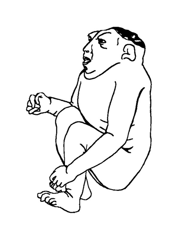 | 600 × 765 (40 KB) | Z8600021 (talk | contribs) | {{Nanagas1925 figures}} |
You cannot overwrite this file.