File:Mousetounge histologicalstain.jpg: Difference between revisions
No edit summary |
No edit summary |
||
| (One intermediate revision by the same user not shown) | |||
| Line 1: | Line 1: | ||
{Student image||2012} | {Student image||2012} | ||
== Mouse Tongue, Histological Stain == | |||
Histological examination is a technique used to examine specimen features in a thorough, specific manner. Structures under histological veiw are easily identifiable and differences and abnormalities can easily be noted. This image was uploaded not to highlight any aspect of taste or tongue development, yet to give an example of contempory techniques used in research. | Histological examination is a technique used to examine specimen features in a thorough, specific manner. Structures under histological veiw are easily identifiable and differences and abnormalities can easily be noted. This image was uploaded not to highlight any aspect of taste or tongue development, yet to give an example of contempory techniques used in research. | ||
== Original text == | |||
Fig 2 Histological findings of tongue in mice treated with 4NQO during 16 weeks. a Normal tongue with no histopathological changes 24 weeks after starting the experiment. b Mild dysplasia after 24 weeks. c Moderate dysplasia after 28 weeks. d Severe dysplasia after 28 weeks. e Invasive squamous cell carcinoma after 32 weeks. Original magnification ×10 | Fig 2 Histological findings of tongue in mice treated with 4NQO during 16 weeks. a Normal tongue with no histopathological changes 24 weeks after starting the experiment. b Mild dysplasia after 24 weeks. c Moderate dysplasia after 28 weeks. d Severe dysplasia after 28 weeks. e Invasive squamous cell carcinoma after 32 weeks. Original magnification ×10 | ||
by Schoop, Remilio A. L.; Noteborn, Mathieu H. M.; Baatenburg de Jong, Robert J. Journal: Journal of Molecular Histology Vol. 40 Issue 3 DOI: 10.1007/s10735-009-9228-z Published: 2009-10-27 Institution(s): Leiden University Medical Center, Leiden University, Erasmus Medical Center | by Schoop, Remilio A. L.; Noteborn, Mathieu H. M.; Baatenburg de Jong, Robert J. Journal: Journal of Molecular Histology Vol. 40 Issue 3 DOI: 10.1007/s10735-009-9228-z Published: 2009-10-27 Institution(s): Leiden University Medical Center, Leiden University, Erasmus Medical Center | ||
http://www.springerimages.com/Images/LifeSciences/1-10.1007_s10735-009-9228-z-1 | |||
== Copyright Information == | |||
This image is copyrighted by The Author(s). This image is published with open access and made available for noncommercial purposes. For more information on what you are allowed to do with this image, please see the Creative Commons pages. | This image is copyrighted by The Author(s). This image is published with open access and made available for noncommercial purposes. For more information on what you are allowed to do with this image, please see the Creative Commons pages. | ||
You are free: | |||
to Share — to copy, distribute and transmit the work to Remix — to adapt the work | |||
Under the following conditions: | |||
Attribution — You must attribute the work in the manner specified by the author or licensor (but not in any way that suggests that they endorse you or your use of the work). | |||
Attribute this work: | |||
What does "Attribute this work" mean? | |||
The page you came from contained embedded licensing metadata, including how the creator wishes to be attributed for re-use. You can use the HTML here to cite the work. Doing so will also include metadata on your page so that others can find the original work as well. Noncommercial — You may not use this work for commercial purposes. | |||
Latest revision as of 11:19, 3 October 2012
{Student image||2012}
Mouse Tongue, Histological Stain
Histological examination is a technique used to examine specimen features in a thorough, specific manner. Structures under histological veiw are easily identifiable and differences and abnormalities can easily be noted. This image was uploaded not to highlight any aspect of taste or tongue development, yet to give an example of contempory techniques used in research.
Original text
Fig 2 Histological findings of tongue in mice treated with 4NQO during 16 weeks. a Normal tongue with no histopathological changes 24 weeks after starting the experiment. b Mild dysplasia after 24 weeks. c Moderate dysplasia after 28 weeks. d Severe dysplasia after 28 weeks. e Invasive squamous cell carcinoma after 32 weeks. Original magnification ×10 by Schoop, Remilio A. L.; Noteborn, Mathieu H. M.; Baatenburg de Jong, Robert J. Journal: Journal of Molecular Histology Vol. 40 Issue 3 DOI: 10.1007/s10735-009-9228-z Published: 2009-10-27 Institution(s): Leiden University Medical Center, Leiden University, Erasmus Medical Center
http://www.springerimages.com/Images/LifeSciences/1-10.1007_s10735-009-9228-z-1
Copyright Information
This image is copyrighted by The Author(s). This image is published with open access and made available for noncommercial purposes. For more information on what you are allowed to do with this image, please see the Creative Commons pages.
You are free: to Share — to copy, distribute and transmit the work to Remix — to adapt the work
Under the following conditions: Attribution — You must attribute the work in the manner specified by the author or licensor (but not in any way that suggests that they endorse you or your use of the work).
Attribute this work:
What does "Attribute this work" mean?
The page you came from contained embedded licensing metadata, including how the creator wishes to be attributed for re-use. You can use the HTML here to cite the work. Doing so will also include metadata on your page so that others can find the original work as well. Noncommercial — You may not use this work for commercial purposes.
File history
Click on a date/time to view the file as it appeared at that time.
| Date/Time | Thumbnail | Dimensions | User | Comment | |
|---|---|---|---|---|---|
| current | 18:06, 1 October 2012 | 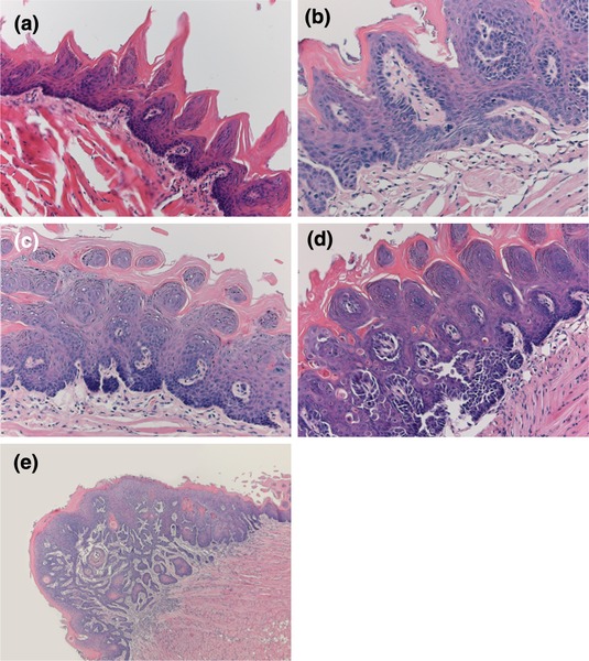 | 535 × 600 (138 KB) | Z3330795 (talk | contribs) | (Student Image) Histological examination is a technique used to examine specimen features in a thorough, specific manner. Structures under histological veiw are easily identifiable and differences and abnormalities can easily be noted. This image was uplo |
You cannot overwrite this file.
File usage
The following file is a duplicate of this file (more details):
The following 2 pages use this file: