File:Mouse telencephalon radial glia model.jpg: Difference between revisions
mNo edit summary |
|||
| Line 19: | Line 19: | ||
* The laminar organisation of both basal progenitors and neurons is disrupted following the loss of functional Dicer. | * The laminar organisation of both basal progenitors and neurons is disrupted following the loss of functional Dicer. | ||
{{Mouse E days}} | |||
===Reference=== | ===Reference=== | ||
Revision as of 12:39, 28 June 2018
Mouse Telencephalon Radial Glia Model
Changes to radial progenitors and their progeny in Dicer1-/- telencephalon.
- At E10.25 the telencephalon comprises the neuroepithelial stem cells “1” which express stem cell markers Sox2, Musashi, CD133.
- Expression of these proteins is maintained throughout their undifferentiated state. By E11.5, the neuroepithelial stem cells establish the radial glia “2”.
- Basal progenitors “3” and neurons “4, 5” are two classes of progeny generated by radial glia around E11.5.
- Dicer deficient neuroepithelium does not establish the appropriate molecular signature of radial glia at ME11.5, which normally strongly express Nestin and Sox9 proteins.
- The proportion of Tbr2 positive basal progenitors is increased in Dicer1-/- telencephalon while the proportions of early postmitotic neurons labelled with TuJ1 and HuC/D or differentiated Tbr1 positive neurons are unchanged.
- The laminar organisation of both basal progenitors and neurons is disrupted following the loss of functional Dicer.
- Mouse Stages: E1 | E2.5 | E3.0 | E3.5 | E4.5 | E5.0 | E5.5 | E6.0 | E7.0 | E7.5 | E8.0 | E8.5 | E9.0 | E9.5 | E10 | E10.5 | E11 | E11.5 | E12 | E12.5 | E13 | E13.5 | E14 | E14.5 | E15 | E15.5 | E16 | E16.5 | E17 | E17.5 | E18 | E18.5 | E19 | E20 | Timeline | About timed pregnancy
| Carnegie | Stage | |||||||||||||||||||||||
| Human | Days | 1 | 2-3 | 4-5 | 5-6 | 7-12 | 13-15 | 15-17 | 17-19 | 20 | 22 | 24 | 28 | 30 | 33 | 36 | 40 | 42 | 44 | 48 | 52 | 54 | 55 | 58 |
| Mouse | Days | 1 | 2 | 3 | E4.5 | E5.0 | E6.0 | E7.0 | E8.0 | E9.0 | E9.5 | E10 | E10.5 | E11 | E11.5 | E12 | E12.5 | E13 | E13.5 | E14 | E14.5 | E15 | E15.5 | E16 |
| Rat | Days | 1 | 3.5 | 4-5 | 5 | 6 | 7.5 | 8.5 | 9 | 10.5 | 11 | 11.5 | 12 | 12.5 | 13 | 13.5 | 14 | 14.5 | 15 | 15.5 | 16 | 16.5 | 17 | 17.5 |
| Note these Carnegie stages are only approximate day timings for average of embryos. Links: Carnegie Stage Comparison | ||||||||||||||||||||||||
| ||||||||||||||||||||||||
| Timeline Links: human timeline | mouse timeline | mouse detailed timeline | chicken timeline | rat timeline | Medaka | Category:Timeline |
Reference
Nowakowski TJ, Mysiak KS, Pratt T & Price DJ. (2011). Functional dicer is necessary for appropriate specification of radial glia during early development of mouse telencephalon. PLoS ONE , 6, e23013. PMID: 21826226 DOI.
Copyright
© 2011 Nowakowski et al. This is an open-access article distributed under the terms of the Creative Commons Attribution License, which permits unrestricted use, distribution, and reproduction in any medium, provided the original author and source are credited.
Original file name: Figure 6. Journal.pone.0023013.g006.jpg dpi:10.1371/journal.pone.0023013.g006
Cite this page: Hill, M.A. (2024, April 19) Embryology Mouse telencephalon radial glia model.jpg. Retrieved from https://embryology.med.unsw.edu.au/embryology/index.php/File:Mouse_telencephalon_radial_glia_model.jpg
- © Dr Mark Hill 2024, UNSW Embryology ISBN: 978 0 7334 2609 4 - UNSW CRICOS Provider Code No. 00098G
File history
Click on a date/time to view the file as it appeared at that time.
| Date/Time | Thumbnail | Dimensions | User | Comment | |
|---|---|---|---|---|---|
| current | 12:34, 2 September 2011 | 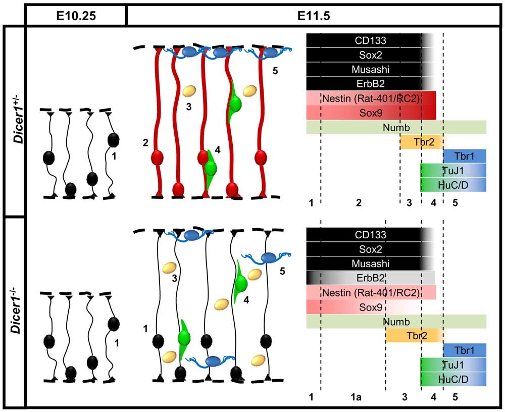 | 1,000 × 816 (80 KB) | S8600021 (talk | contribs) | ==Mouse Telencephalon Radial Glia Model== Changes to radial progenitors and their progeny in Dicer1-/- telencephalon. * At E10.25 the telencephalon comprises the neuroepithelial stem cells “1” which express stem cell markers Sox2, Musashi, CD133. * |
You cannot overwrite this file.
File usage
There are no pages that use this file.