File:Mouse renal podocyte EM01.jpg

Original file (1,000 × 1,338 pixels, file size: 366 KB, MIME type: image/jpeg)
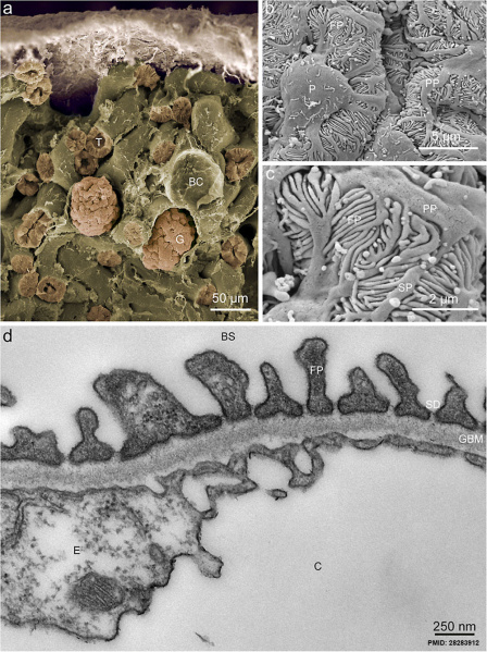
Mouse Glomerular Ultrastructure
Reference
<pubmed>28283912</pubmed>
New structural insights into podocyte biology Cell Tissue Res. 2017 Mar 10. doi: 10.1007/s00441-017-2590-3.
Copyright
Grahammer F
© The Author(s) 2017 Open Access This article is distributed under the terms of the Creative Commons Attribution 4.0 International License (http://creativecommons.org/licenses/by/4.0/), which permits unrestricted use, distribution, and reproduction in any medium, provided you give appropriate credit to the original author(s) and the source, provide a link to the Creative Commons license, and indicate if changes were made. PMID added and image resized.
Cite this page: Hill, M.A. (2024, April 25) Embryology Mouse renal podocyte EM01.jpg. Retrieved from https://embryology.med.unsw.edu.au/embryology/index.php/File:Mouse_renal_podocyte_EM01.jpg
- © Dr Mark Hill 2024, UNSW Embryology ISBN: 978 0 7334 2609 4 - UNSW CRICOS Provider Code No. 00098G
File history
Click on a date/time to view the file as it appeared at that time.
| Date/Time | Thumbnail | Dimensions | User | Comment | |
|---|---|---|---|---|---|
| current | 13:37, 28 March 2017 | | 1,000 × 1,338 (366 KB) | Z8600021 (talk | contribs) | ==Mouse Glomerular Ultrastructure== PMID 28283912 |
You cannot overwrite this file.
File usage
The following page uses this file: