File:Mouse pronuclei 02.jpg
Original file (800 × 801 pixels, file size: 58 KB, MIME type: image/jpeg)
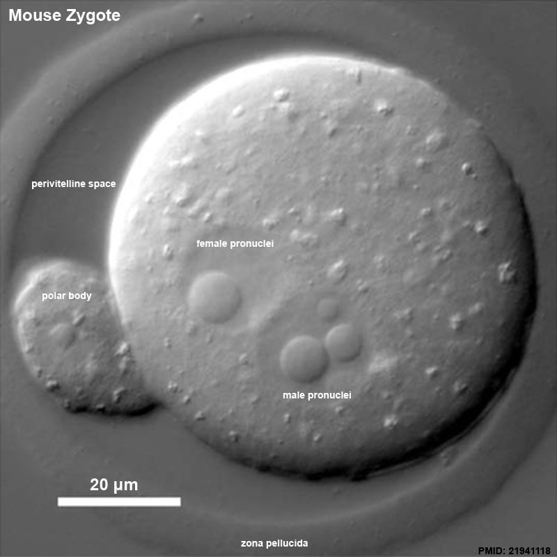
Mouse Pronuclei
DIC image of a living mouse egg shortly before fusion of the two pronuclei. In this focal plane the female pronucleus displays a single large nuclear body, whereas the slightly larger male pronucleus shows one small and two large bodies. The polar body nucleus also appears to contain a small nuclear body. Note the superficial similarity to the induced nuclear bodies of Drosophila.
Reference
<pubmed>21941118</pubmed>| Nucleus
Copyright
© 2011 Landes Bioscience.
To secure the rights to reuse materials from this paper, please visit the The Copyright Clearance Center. This is an open-access article licensed under a Creative Commons Attribution-NonCommercial 3.0 Unported License. The article may be redistributed, reproduced, and reused for non-commercial purposes, provided the original source is properly cited.
http://creativecommons.org/licenses/by-nc/3.0/
https://www.landesbioscience.com/article/17250/full_text/#load/figures/text-15/image-fig-F8
nucl0205_0403_fig008.jpg
File history
Click on a date/time to view the file as it appeared at that time.
| Date/Time | Thumbnail | Dimensions | User | Comment | |
|---|---|---|---|---|---|
| current | 19:17, 5 August 2014 | | 800 × 801 (58 KB) | Z8600021 (talk | contribs) | ==Mouse Pronuclei== DIC image of a living mouse egg shortly before fusion of the two pronuclei. In this focal plane the female pronucleus displays a single large nuclear body, whereas the slightly larger male pronucleus shows one small and two large b... |
You cannot overwrite this file.
File usage
The following 3 pages use this file: