File:Mouse oocytes in vitro.png: Difference between revisions
No edit summary |
No edit summary |
||
| Line 1: | Line 1: | ||
==Growth of Mouse Oocytes to Maturity from Premeiotic Germ Cells In Vitro== | |||
(A–E) Example of 2-cell embryo, 4-cell embryo, morula and blastocyst developed from oocytes generated in vitro in the presence of ActA. Left, phase contrast microscopy observations; right, Hoechst staining of the cell nuclei. | (A–E) Example of 2-cell embryo, 4-cell embryo, morula and blastocyst developed from oocytes generated in vitro in the presence of ActA. Left, phase contrast microscopy observations; right, Hoechst staining of the cell nuclei. | ||
Latest revision as of 22:38, 7 August 2012
Growth of Mouse Oocytes to Maturity from Premeiotic Germ Cells In Vitro
(A–E) Example of 2-cell embryo, 4-cell embryo, morula and blastocyst developed from oocytes generated in vitro in the presence of ActA. Left, phase contrast microscopy observations; right, Hoechst staining of the cell nuclei.
Reference
1. Zhang Z-P, Liang G-J, Zhang X-F, Zhang G-L, Chao H-H, et al. (2012) Growth of Mouse Oocytes to Maturity from Premeiotic Germ Cells In Vitro. PLoS ONE 7(7): e41771. doi:10.1371/journal.pone.0041771
Copyright: © 2012 Shen et al. This is an open-access article distributed under the terms of the Creative Commons Attribution License, which permits unrestricted use, distribution, and reproduction in any medium, provided the original author and source are credited.
- Note - This image was originally uploaded as part of an undergraduate science student project and may contain inaccuracies in either description or acknowledgements. Students have been advised in writing concerning the reuse of content and may accidentally have misunderstood the original terms of use. If image reuse on this non-commercial educational site infringes your existing copyright, please contact the site editor for immediate removal.
File history
Click on a date/time to view the file as it appeared at that time.
| Date/Time | Thumbnail | Dimensions | User | Comment | |
|---|---|---|---|---|---|
| current | 22:21, 7 August 2012 | 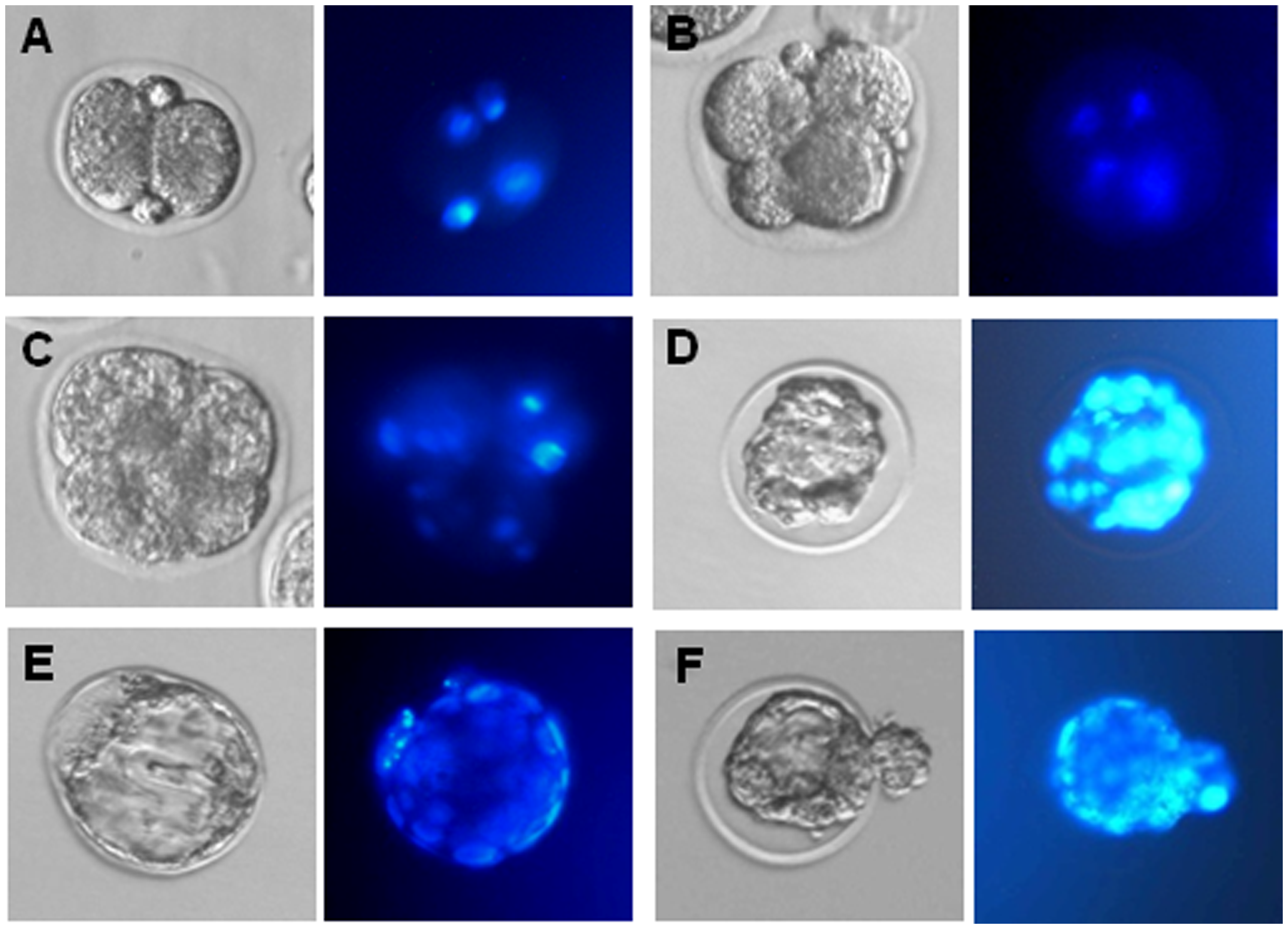 | 3,439 × 2,481 (4.19 MB) | Z3330986 (talk | contribs) | (A–E) Example of 2-cell embryo, 4-cell embryo, morula and blastocyst developed from oocytes generated in vitro in the presence of ActA. Left, phase contrast microscopy observations; right, Hoechst staining of the cell nuclei. |
You cannot overwrite this file.
File usage
The following page uses this file: