File:Mouse inner cell mass cell types 01.jpg: Difference between revisions
No edit summary |
|||
| Line 8: | Line 8: | ||
'''(C)''' Lineage tree from representative embryo. All cells were traced to the early 32-cell blastocyst; then inside cells were traced to late blastocyst. A cell was defined as occupying an inside position by the orientation of the cell division that generated it, by its enclosure from the outside environment by neighboring cells, and by continued analysis of its position as development progressed. Allocation to trophectoderm (TE), EPI, or PE and apoptosis (A) are indicated. | '''(C)''' Lineage tree from representative embryo. All cells were traced to the early 32-cell blastocyst; then inside cells were traced to late blastocyst. A cell was defined as occupying an inside position by the orientation of the cell division that generated it, by its enclosure from the outside environment by neighboring cells, and by continued analysis of its position as development progressed. Allocation to trophectoderm (TE), EPI, or PE and apoptosis (A) are indicated. | ||
:'''Links:''' [[Mouse Development]] | [[Morula]] | [[Blastocyst]] | |||
===Reference=== | ===Reference=== | ||
Revision as of 06:27, 13 November 2011
Mouse inner cell mass cell types
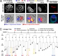
Origin and formation of the first two distinct cell types of the inner cell mass in the mouse embryo using live cell imaging and tracking.
(A) GFP-GPI expression from E.2.5 to E4.5. Deconvolved fluorescence and differential interference contrast time-lapse images overlaid with Simi BioCell cell-tracking spheres.
(B) Embryos stained to reveal Gata4-positive cells adjacent to mature blastocyst cavity confirming normal development during each imaging session. Gata4-positive cells were present in a one-cell-thick surface layer.
(C) Lineage tree from representative embryo. All cells were traced to the early 32-cell blastocyst; then inside cells were traced to late blastocyst. A cell was defined as occupying an inside position by the orientation of the cell division that generated it, by its enclosure from the outside environment by neighboring cells, and by continued analysis of its position as development progressed. Allocation to trophectoderm (TE), EPI, or PE and apoptosis (A) are indicated.
- Links: Mouse Development | Morula | Blastocyst
Reference
<pubmed>20308546</pubmed>| PMC2852013 | PNAS
Department of Physiology, Wellcome Trust/Cancer Research UK Gurdon Institute, University of Cambridge, Cambridge CB2 3EH, United Kingdom.
Copyright
Proceedings National Academy of Sciences (PNAS) Liberalization of PNAS copyright policy: Noncommercial use freely allowed Note original Author should be contacted for permission to reuse for Educational purposes. See also PNAS Author Rights and Permission FAQs
- Cozzarelli NR, Fulton KR, Sullenberger DM. Liberalization of PNAS copyright policy: noncommercial use freely allowed. Proc Natl Acad Sci U S A. 2004 Aug 24;101(34):12399. PMID15314225 "Our guiding principle is that, while PNAS retains copyright, anyone can make noncommercial use of work in PNAS without asking our permission, provided that the original source is cited."
Fig. 1. http://www.pnas.org/content/107/14/6364/F1.large.jpg (Original image scaled and text from figure legend)
File history
Click on a date/time to view the file as it appeared at that time.
| Date/Time | Thumbnail | Dimensions | User | Comment | |
|---|---|---|---|---|---|
| current | 06:22, 13 November 2011 |  | 820 × 800 (128 KB) | S8600021 (talk | contribs) | ==Mouse inner cell mass cell types== Origin and formation of the first two distinct cell types of the inner cell mass in the mouse embryo Live cell imaging and tracking. (A) GFP-GPI expression from E.2.5 to E4.5. Deconvolved fluorescence and differentia |
You cannot overwrite this file.
File usage
The following page uses this file: