File:Mouse heart E9.5.jpg
Mouse_heart_E9.5.jpg (600 × 558 pixels, file size: 31 KB, MIME type: image/jpeg)
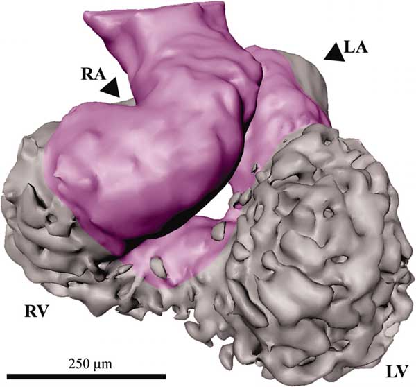
An ED 9.5 lumen. The primary heart tube is shown in purple, and the left and right ventricles (LV and RV) and mostly obscured left and right atria (LA and RA) are depicted in gray.
These reconstructions support the ballooning-heart model (3, 5) in which the chambers are thought to "balloon" out of the primary tube, as opposed to the textbook model of heart development, the so-called segmental model.
Image (used with permission) from the paper Soufan AT, Ruijter JM, van den Hoff MJ, de Boer PA, Hagoort J, Moorman AF. Three-dimensional reconstruction of gene expression patterns during cardiac development. Physiol Genomics. 2003 May 13;13(3):187-95. (Physiol Genomics paper)
MouseheartF6.jpg
http://physiolgenomics.physiology.org/cgi/content-nw/full/13/3/187/F6
File history
Click on a date/time to view the file as it appeared at that time.
| Date/Time | Thumbnail | Dimensions | User | Comment | |
|---|---|---|---|---|---|
| current | 20:44, 16 August 2009 | | 600 × 558 (31 KB) | S8600021 (talk | contribs) | Image (used with permission) from the paper Soufan AT, Ruijter JM, van den Hoff MJ, de Boer PA, Hagoort J, Moorman AF. Three-dimensional reconstruction of gene expression patterns during cardiac development. Physiol Genomics. 2003 May 13;13(3):187-95. (Ph |
You cannot overwrite this file.
File usage
The following 4 pages use this file: