File:Mouse embryo vascular.png: Difference between revisions
No edit summary |
m (→Reference) |
||
| (11 intermediate revisions by 2 users not shown) | |||
| Line 1: | Line 1: | ||
==Surface | ==Surface Renderings of Embryonic Vascular Structures== | ||
(A) Reconstructed FDR-deconvolution OPT data of the 19 somite embryo is shown as a surface rendered object. | (A) Reconstructed FDR-deconvolution OPT data of the 19 somite embryo is shown as a surface rendered object. | ||
| Line 11: | Line 11: | ||
(E) Segmentation of the data allows selective display of labelled structures. Exclusion of the unsegmented data provides better analysis of the ICAs and the pharyngeal arch arteries. | (E) Segmentation of the data allows selective display of labelled structures. Exclusion of the unsegmented data provides better analysis of the ICAs and the pharyngeal arch arteries. | ||
:'''Links:''' [[Movie - Mouse Cephalic Plexus]] | [[Cardiovascular System Development]] | [[Mouse Development]] | |||
All scale bars represent 100 microns. | All scale bars represent 100 microns. | ||
'''ACV''' - anterior cardinal vein | ===Legend=== | ||
* '''ACV''' - anterior cardinal vein | |||
'''CCV''' - common cardinal vein | * '''CCV''' - common cardinal vein | ||
* '''DA''' - dorsal aorta | |||
'''DA''' - dorsal aorta | * '''DLAV''' - dorsal longitudinal anastomotical vessel | ||
* '''ISA''' - intersomitic artery | |||
'''DLAV''' - dorsal longitudinal anastomotical vessel | * '''ISV''' - intersomitic vein | ||
* '''OA''' - omphalomesenteric artery | |||
'''ISA''' - intersomitic artery | * '''OV''' - omphalomesenteric vein | ||
* '''PCV''' - posterior cardinal vein | |||
* '''PNVP''' - perineual vascular plexus | |||
* '''UA''' - umbilical artery | |||
* '''UV''' - umbilical vein | |||
===Staging=== | |||
* [[Mouse_Stages#Theiler_Stage_14|Theiler Stage 14]] Embryonic age = 9 dpc (range 8.5-9.75 dpc) | |||
* Looks like equivalent Carnegie Stage in humans = 12 | |||
===Reference=== | |||
{{#pmid:18682734}} | |||
====Copyright==== | |||
© 2008 Walls et al. | |||
This is an open-access article distributed under the terms of the Creative Commons Attribution License, which permits unrestricted use, distribution, and reproduction in any medium, provided the original author and source are credited. | |||
Original File name: Figure 2. Journal.pone.0002853.g002.png | Original File name: Figure 2. Journal.pone.0002853.g002.png | ||
{{Footer}} | |||
[[Category:Heart]] [[Category:Cardiovascular]] [[Category:Mouse]] | |||
[[Category: | |||
Latest revision as of 13:34, 18 July 2019
Surface Renderings of Embryonic Vascular Structures
(A) Reconstructed FDR-deconvolution OPT data of the 19 somite embryo is shown as a surface rendered object.
(B) The surface rendered object can be zoomed in to any magnification, as in this magnified image of the vasculature in the mouse head.
(C) The surface rendering can also be rotated so that it can be viewed from any angle. Viewing the rendering from the left side reveals structures in the heart (arrow) that are obscured by the tail in (A).
(D) The 3D data can also be segmented as described in Materials and Methods. The DA, heart and ICAs are labelled yellow, the UV dark pink, and the unsegmented vasculature blue.
(E) Segmentation of the data allows selective display of labelled structures. Exclusion of the unsegmented data provides better analysis of the ICAs and the pharyngeal arch arteries.
All scale bars represent 100 microns.
Legend
- ACV - anterior cardinal vein
- CCV - common cardinal vein
- DA - dorsal aorta
- DLAV - dorsal longitudinal anastomotical vessel
- ISA - intersomitic artery
- ISV - intersomitic vein
- OA - omphalomesenteric artery
- OV - omphalomesenteric vein
- PCV - posterior cardinal vein
- PNVP - perineual vascular plexus
- UA - umbilical artery
- UV - umbilical vein
Staging
- Theiler Stage 14 Embryonic age = 9 dpc (range 8.5-9.75 dpc)
- Looks like equivalent Carnegie Stage in humans = 12
Reference
Walls JR, Coultas L, Rossant J & Henkelman RM. (2008). Three-dimensional analysis of vascular development in the mouse embryo. PLoS ONE , 3, e2853. PMID: 18682734 DOI.
Copyright
© 2008 Walls et al. This is an open-access article distributed under the terms of the Creative Commons Attribution License, which permits unrestricted use, distribution, and reproduction in any medium, provided the original author and source are credited.
Original File name: Figure 2. Journal.pone.0002853.g002.png
Cite this page: Hill, M.A. (2024, April 25) Embryology Mouse embryo vascular.png. Retrieved from https://embryology.med.unsw.edu.au/embryology/index.php/File:Mouse_embryo_vascular.png
- © Dr Mark Hill 2024, UNSW Embryology ISBN: 978 0 7334 2609 4 - UNSW CRICOS Provider Code No. 00098G
File history
Click on a date/time to view the file as it appeared at that time.
| Date/Time | Thumbnail | Dimensions | User | Comment | |
|---|---|---|---|---|---|
| current | 10:07, 15 August 2009 | 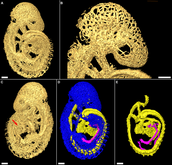 | 600 × 576 (518 KB) | S8600021 (talk | contribs) | Figure 2. Surface renderings of embryonic vascular structures. (A) Reconstructed FDR-deconvolution OPT data of the 19 somite embryo is shown as a surface rendered object. (B) The surface rendered object can be zoomed in to any magnification, as in this |
You cannot overwrite this file.