File:Mouse diencephalon territories 01.jpg
Mouse_diencephalon_territories_01.jpg (490 × 343 pixels, file size: 81 KB, MIME type: image/jpeg)
Mouse diencephalon territories
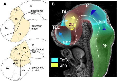
(A) Schematic representations of a lateral view of E10.5 mouse neural tube where diencephalic territories (yellow) have been represented following columnar (above) and prosomeric (below) models.
(B) Scanning electron microscope image showing a lateral view of E10.5 mouse neural tube where the main neural segments and planar secondary organizers are represented.
Abbreviations: anr, anterior neural ridge; Di, diencephalon; Eth, epithalamus; Hy, hypothalamus; IsO, isthmic organizer; M, mesencephalon; PT, pretectum; Rh, rhombencephalon; Tel, telencephalon; Th, thalamus; VTh (PTh), ventral thalamus (prethalamus); ZLI, zona limitans intrathalamica.
Reference
Martinez-Ferre A & Martinez S. (2012). Molecular regionalization of the diencephalon. Front Neurosci , 6, 73. PMID: 22654731 DOI.
Copyright
© 2012 Martinez-Ferre and Martinez. This is an open-access article distributed under the terms of the Creative Commons Attribution Non Commercial License, which permits non-commercial use, distribution, and reproduction in other forums, provided the original authors and source are credited.
Cite this page: Hill, M.A. (2024, April 18) Embryology Mouse diencephalon territories 01.jpg. Retrieved from https://embryology.med.unsw.edu.au/embryology/index.php/File:Mouse_diencephalon_territories_01.jpg
- © Dr Mark Hill 2024, UNSW Embryology ISBN: 978 0 7334 2609 4 - UNSW CRICOS Provider Code No. 00098G
File history
Click on a date/time to view the file as it appeared at that time.
| Date/Time | Thumbnail | Dimensions | User | Comment | |
|---|---|---|---|---|---|
| current | 14:10, 23 November 2019 | | 490 × 343 (81 KB) | Z8600021 (talk | contribs) | (A) Schematic representations of a lateral view of {{ME10.5}} mouse neural tube where diencephalic territories (yellow) have been represented following columnar (above) and prosomeric (below) models. (B) Scanning electron microscope image showing a l... |
You cannot overwrite this file.
File usage
The following page uses this file: