File:Mouse cerebellum connections 01.jpg: Difference between revisions
Mouse_cerebellum_connections_01.jpg (470 × 600 pixels, file size: 44 KB, MIME type: image/jpeg)
| Line 8: | Line 8: | ||
Abbreviations: AMG, amygdala; BG, basal ganglia; ECN, external cuneate nucleus; HIP, hippocampus; HYP; hypothalamus; IO, inferior olive; LC, locus coeruleus; PAG, periaqueductal gray; PN, pontine nuclei; RET, reticular nucleus; RN, red nucleus; SC, spinal cord; SUP, superior colliculi; TH, thalamus; VN, vestibular nuclei. | Abbreviations: AMG, amygdala; BG, basal ganglia; ECN, external cuneate nucleus; HIP, {{hippocampus}}; HYP; {{hypothalamus}}; IO, inferior olive; LC, locus coeruleus; PAG, periaqueductal gray; PN, pontine nuclei; RET, reticular nucleus; RN, red nucleus; SC, spinal cord; SUP, superior colliculi; TH, thalamus; VN, vestibular nuclei. | ||
| Line 25: | Line 25: | ||
[[Category:Mouse]][[Category:Cerebellum]][[Category:Cartoon]] | [[Category:Mouse]][[Category:Cerebellum]][[Category:Cartoon]] | ||
[[Category:Hippocampus]] | |||
Latest revision as of 11:19, 17 January 2020
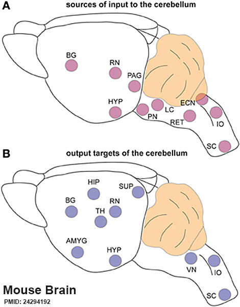
Mouse Cerebellum Connections
The cerebellum is extensively connected to the brain and spinal cord.
A - Schematic representation of brain regions that send input to the cerebellum.
B - Schematic representation of the regions that receive information from the cerebellum. Note that the TH is a major relay station for cerebellar input to the cortex while the PN is the primary gateway for cerebral cortical input to the cerebellum.
Abbreviations: AMG, amygdala; BG, basal ganglia; ECN, external cuneate nucleus; HIP, hippocampus; HYP; hypothalamus; IO, inferior olive; LC, locus coeruleus; PAG, periaqueductal gray; PN, pontine nuclei; RET, reticular nucleus; RN, red nucleus; SC, spinal cord; SUP, superior colliculi; TH, thalamus; VN, vestibular nuclei.
- Links: cerebellum | mouse
Reference
Reeber SL, Otis TS & Sillitoe RV. (2013). New roles for the cerebellum in health and disease. Front Syst Neurosci , 7, 83. PMID: 24294192 DOI.
Copyright
© 2013 Reeber, Otis and Sillitoe. This is an open-access article distributed under the terms of the Creative Commons Attribution License (CC BY). The use, distribution or reproduction in other forums is permitted, provided the original author(s) or licensor are credited and that the original publication in this journal is cited, in accordance with accepted academic practice. No use, distribution or reproduction is permitted which does not comply with these terms.
Fig 3 Fnsys-07-00083-g003.jpg adjusted in size and labelling.
Cite this page: Hill, M.A. (2024, April 24) Embryology Mouse cerebellum connections 01.jpg. Retrieved from https://embryology.med.unsw.edu.au/embryology/index.php/File:Mouse_cerebellum_connections_01.jpg
- © Dr Mark Hill 2024, UNSW Embryology ISBN: 978 0 7334 2609 4 - UNSW CRICOS Provider Code No. 00098G
File history
Click on a date/time to view the file as it appeared at that time.
| Date/Time | Thumbnail | Dimensions | User | Comment | |
|---|---|---|---|---|---|
| current | 15:06, 16 February 2016 | | 470 × 600 (44 KB) | Z8600021 (talk | contribs) | ==Mouse Cerebellum Connections== The cerebellum is extensively connected to the brain and spinal cord. (A) Schematic representation of brain regions that send input to the cerebellum. (B) Schematic representation of the regions that receive inform... |
You cannot overwrite this file.
File usage
The following page uses this file: